 |
| About Bioline | All Journals | Testimonials | Membership | News |
|
||||||
|
||||||
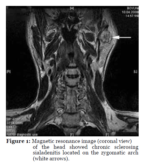
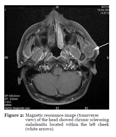
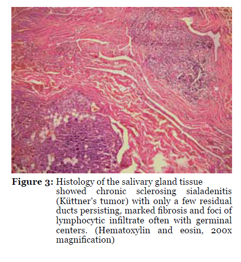
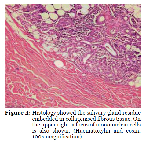
Malaysian Journal of Medical Sciences, Vol. 17, No. 3, 2010, pp. 57-61 Case Report Chronic Sclerosing Sialadenitis (Küttner’s tumour) of the Parotid Gland Güçlü Kaan Beriat1 , Şefik Halit Akmansu1, Sinan Kocatürk1, Ömür Ataoğlu2 1Department of Otolaryngology, Faculty of Medicine, Ufuk University, No:86, Konya Avenue, 06520 Balgat, Ankara, Turkey Correspondence: Güçlü Kaan BeriatMD (Ufuk University)Department of OtolaryngologyUfuk University No:86, Konya Avenue 06520 BalgatAnkara, Turkey, Tel: +90 0532 476 97 20 (GSM), +90 0312 204 41 75 (Office) Email: beriat4@gmail.com, kberiat@hotmail.com Submitted: 4 Dec 2009 Code Number: mj10049 Abstract Chronic sclerosing sialadenitis is a chronic inflammatory salivary gland disease. Küttner reported 4 cases of submandibular gland lesions for the first time in 1896. Chronic sclerosing sialadenitis is a very rare inflammatory lesion of the parotid gland and cannot be easily distinguished from salivary malignant masses. We reported a 28-year-old male with a painful parotid tumour, which grew slowly for 4 years. Keywords: chronic illness, inflammation, parotid gland, sclerosis, sialadenitis Introduction A series of patients with unilateral, hard, tumour-like masses of the submandibular gland were diagnosed with chronic sclerosing sialadenitis by Küttner in 1896 (1). This disease is clinically similar to salivary gland neoplasms and is classified as a tumour-like lesion of the salivary glands by the World Health Organization (2). Chronic sclerosing sialadenitis is clinically characterised by a firm, relatively painful swelling of one of the submandibular glands. This disorder is characterised by plasmocytic and lymphocytic periductal infiltrate eventually leading to encasement of ducts with thick fibrous tissue (3). Histologically, chronic sclerosing sialadenitis is characterised by periductal sclerosis, acinar atrophy, and infiltration of the gland by lymphocytes, which some studies have recognised as predominantly activated B cells with a subpopulation of helper-inducer T cells. The distribution pattern of these lymphocytes suggested that the response was immunological. However, sialoliths and mucous plugs were found in 29% to 83% of the lesions. This association was meaningful for some authors, such that it was considered in favour of a cause and result relationship. Other possible aetiologies of chronic sclerosing sialadenitis are ascending bacterial infections of the oral cavity and duct obstruction by foreign bodies. Case Report A 28-year-old male was investigated at the Otolaryngology Outpatient Department. He first noted the mass 4 years earlier. Physical examination revealed a tender, hard and fixed, 3 x 2 cm mass at the angle of the left maxillary arch, and it seemed to be attached to underlying structures. No other masses or adenopathy were noted in the head or neck. No related events were present in the patient’s medical history. He reported no other symptoms or complaints. His facial nerve function was intact. A sonographically guided fine-needle biopsy was performed. Cytology revealed only lymphocytes and other blood elements; however, no glandular epithelial cells were present. Magnetic resonance imaging detected a 3 x 2.5 x 2 cm, smooth-surfaced, multilobular mass in the left parotid gland. This mass had a heterogenic opaque appearance after intravenous contrast injection. No pathologic nodes were identified (Figures 1 and 2). Left total parotidectomy was conducted with mass excision and preservation of the facial nerve. The specimen collected for pathological examination measured 2.5 cm at its longest diameter. It was a yellow-white in colour, and its cut surface demonstrated a firm consistency. The whole tissue sample was submitted for pathological examination. Pathology revealed chronic sclerosing sialadenitis. The microscopic examination revealed a collagenised fibrous tissue in all sections. Residual salivary gland tissue seemed to be embedded in the fibrous tissue in some areas (Figure 3). In the salivary gland tissue, mononuclear cells, mostly lymphocytes, were also observed (Figure 4). Mononuclear cells were also observed in the fibrous stroma. No evidence of malignancy was observed, and the final diagnosis was chronic sclerosing sialadenitis. Post-operative complications and recurrence were not encountered in the 2-year follow-up. Discussion Chronic sclerosing sialadenitis is an inflammatory process that primarily affects the submandibular gland and presents clinically as a painful swelling. Histologically, chronic sclerosing sialadenitis demonstrates a loss in acinar tissue, dense fibrosis, which is mainly periductal, and lymphocytic infiltration with lymphoid follicle formation in some cases (4). According to Seifert (5), chronic sclerosing sialadenitis may progress through 4 different histological stages:
The origin of chronic sclerosing sialadenitis has been attributed to many etiologic agents. Sialoliths and mucous plugs are found in 29% to 83% of cases, but it is not clear whether the sialoliths are the causes or results of the inflammatory process (2,6,7). In this case, no sialoliths were found. Anything that causes an obstruction of salivary flow or stasis of secretions can lead to acinar cell swelling, necrosis, ductal dilation, and retention of salivary secretions accompanying oedema and inflammatory cell infiltration (8). Salivary gland stones may cause obstruction of salivary flow or stasis of secretions. Seifert and Donath (5) proposed a theory of obstructive electrolyte sialadenitis in which a secretion disorder produces inspissated secretion that obstructs the small ducts, which leads to inflammation, fibrosis, parenchymal atrophy, and an immune reaction of the duct system. Immunohistochemical studies of the lymphoid population in the chronic sclerosing sialadenitis revealed abundant cytotoxic T cells especially near ducts and acini. The B cell reaction was less pronounced and largely restricted to lymph follicles. There was an intimate relationship between the T-cell-dominated inflammatory infiltrate and acinar and duct cells. The monoclonal and oligoclonal populations of cytotoxic T cells and their histopathological behaviours suggested that chronic sclerosing sialadenitis may be the result of an immune process triggered by intraductal epithelial agents. The lymphocytes that infiltrate the epithelial component of chronic sclerosing sialadenitis are mainly B cells and are often characterised by a lack of Bcl-2 expression (9,10). The duration of symptoms before the patient seeks treatment is variable, from 1 month to about 3 decades, and the induration and enlargement often lead clinicians to diagnose chronic sclerosing sialadenitis as a salivary gland neoplasm (7). Possible differential diagnoses of this entity include other benign inflammatory lesions of salivary glands, such as simple chronic sialadenitis, granulomatous sialadenitis, necrotising sialometaplasia, sialolithiasis, radiation effects, an inflammatory pseudotumour, and benign lymphoepithelial lesions. One of the most common causes of chronic inflammation of the salivary glands is related to rheumatoid arthritis, which is consistent with immune pathogenesis (11). Usually, these cases do not demonstrate symptoms related to sialadenitis. However, sialolithiasis is the most common cause of sialadenitis in symptomatic patients. Histologically, various degrees of atrophy, fibrosis, and chronic inflammation are seen, but lymphoplasmocytic periductal infiltrate and fibrous encasement of ducts, which are typical of chronic sclerosing sialadenitis, are not present. The causes of granulomatous sialadenitis range from infections to duct obstruction caused by calculi or malignancies. Granulomas of different types can be present, and a xanthogranulomatous variant has been described (12). Necrotising sialometaplasia occurs mainly in the minor salivary glands of the palate and may be confused with carcinoma. In necrotising sialometaplasia, an ischemic aetiology is thought to produce ulcerating lesions with partial necrosis of glands associated with regeneration and squamous metaplasia of the adjacent ducts. Its localisation and lack of lymphoplasmocytic infiltration differentiates necrotising sialometaplasia from chronic sclerosing sialadenitis. Sialolithiasis can be observed in all major salivary glands, although it is more common in the submandibular gland. Ductal obstruction induces inflammation of the surrounding tissue and acinar atrophy ensures. Radiographic examination may demonstrate a radiopaque mass. Histological examination shows dilated ducts and variable destruction of salivary tissue. The characteristic histological symptoms of chronic sclerosing sialadenitis are not seen. Radiation effects are more frequently seen in the submandibular glands because they are located in the irradiation fields for tumours of the oral cavity. Acinar atrophy, chronic inflammation, and squamous metaplasia are commonly seen. The lymphocyte population is mainly formed by B cells with sparse T cells without the peculiar topographic distribution seen in chronic sclerosing sialadenitis. Inflammatory myofibroblastic tumours are rare inflammatory processes that may occur in the salivary glands primarily as a result of injury. They are composed of lymphocytes, plasma cells, histiocytes, and myofibroblasts, which is in sharp contrast with the monotonous lymphoplasmocytic infiltrate of chronic sclerosing sialadenitis. Benign lymphoepithelial lesions are seen in Mikulicz syndrome, which is a bilateral enlargement of the salivary and lacrimal glands (13). The swelling of the salivary glands is usually symmetric. Mikulicz disease is part of a more complex syndrome known as Sjogren’s syndrome, which also includes keratoconjunctivitis, xerostomia, and rheumatoid arthritis. The main histological features are lymphoid infiltration and epimyoepithelial islands, which consist of solid nests of prominent myoepithelial proliferation infiltrated by a mixed population of B and T lymphocytes. Therefore, the presentation and histological features of Mikulicz disease are different from chronic sclerosing sialadenitis. Chronic sclerosing sialadenitis is a totally benign inflammatory lesion, and until now, there have been no reports of malignancy. In fact, chronic sclerosing sialadenitis has often been removed, and no additional treatment has been required (14). This disease is rare and most commonly occurs in the submandibular gland (15,16). We present a very rare case of chronic sclerosing sialadenitis, which affected the parotid gland as a solitary mass. Authors’ Contributions Provision of patient, collection and assembly of data: OA References
© Copyright 2010 - Malaysian Journal of Medical Science The following images related to this document are available:Photo images[mj10049f1.jpg] [mj10049f4.jpg] [mj10049f3.jpg] [mj10049f2.jpg] |
| |||||||||
