 |
| About Bioline | All Journals | Testimonials | Membership | News |
|
||||||
|
||||||
Malaysian Journal of Medical Sciences, Vol. 18, No. 2, 2011, pp. 40-46 Original Article Influence of Sitting and Prone Lying Positions on Proprioceptive Knee Assessment Score in Early Knee Osteoarthritis Vijay Batra1, Vijai Prakash Sharma1, Meenakshi Batra1, Girdhar Gopal Agarwal2, Vineet Sharma3 1Department of Physical Medicine and
Rehabilitation, Rehabilitation and Artificial Limb Centre, Chhatrapati Shahuji
Maharaj Medical University (Formerly King George Medical University), Dali
Ganj
Bridge, Nabiullah Road, Lucknow: 226018, Uttar Pradesh, India Correspondence: Dr Vijay Batra PhD Occupational Therapy (Chhatrapati Shahuji Maharaj Medical University) Department of Physical Medicine & Rehabilitation Rehabilitation and Artificial Limb Centre Chhatrapati Shahuji Maharaj Medical University (Formerly King George Medical University) Dali Ganj Bridge, Nabiullah Road Lucknow: 226018 Uttar Pradesh, India Tel: +91-9868019077, +91-9044467434 Email: vijaybatra@yahoo.com, vijaybatras@gmail.com Submitted: 9 Mar 2010 Code Number: mj11020 AbstractBackground: Knee proprioception is compromised in
knee osteoarthritis. There are several ways of measuring proprioceptive acuity,
but there is lack of consensus over the ideal testing position. The study aimed
to evaluate the influence of 2 testing positions (sitting versus prone lying)
on proprioceptive knee assessment score in patients with early knee
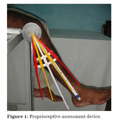
osteoarthritis. Keywords: adaptive behavior, knee, patient positioning, proprioception, osteoarthritis, Introduction Proprioception is the sense of position and movement of the limbs, and it is the result of sensory inputs arising from muscle, skin, and joint structures (1). It can also be defined as the conscious and unconscious awareness of body position, movement, and forces acting on the body, for which accurate sensory input and central integration from peripheral proprioceptors are a must (2). Proprioception contributes to the development of the motor control and plays a major role in the reflex protection of joints against potentially harmful forces (1). The proprioceptive acuity impairment significantly affects neuromusculoskeletal integrity, contributing to pain and functional disability (2–4). Knee proprioception has consistently been reported to be compromised in individuals with knee osteoarthritis (3,5–11). This neuromuscular deficit has been suggested as the major contributing factor to the disease process (7,10–13). However, studies have shown that the impairment of proprioceptive acuity is not exclusively a local result of the disease, and there is a need to study its importance in the development and progression of knee osteoarthritis (10,11). There are several ways of measuring proprioceptive acuity; one of them is the threshold detection of passive movement. However, passive movements do not reflect real life movement or function; proprioceptive functions in healthy and pathological joints are quite variable and there is a lack of correlation between different measurements of proprioception in the knee (14–17). Active assessments by asking the patient to replicate limb position, using active movement, with vision occluded (16) or by reproducing lower limb static loads (15) have been suggested. Generally, proprioceptive assessment of the knee is done in sitting position (16). However, the ideal testing position for proprioceptive assessment of the knee is still debatable. One of the reasons could be that, during assessment, the subject may exhibit an adaptive behaviour to compensate for the loss of proprioceptive acuity by using vision or not relaxing the muscles completely before attempting to replicate limb position. The purpose of the present study was to evaluate the influence of 2 testing positions (sitting and prone lying) on proprioceptive acuity scores (17) in the assessment of early knee osteoarthritis. Subjects and Methods The study involved 70 subjects (22 males and 48 females) with history of knee pain and clinical diagnosis of early knee osteoarthritis, with radiological findings of grade I (33 subjects) and grade II (37 subjects) according to the Kellgren and Lawrence Classification System (18). All patients were between 40 and 60 years of age. Subjects with traumatic knee injury, inflammatory arthritis, metabolic disorder, as well as cardiovascular and psychiatric illnesses were excluded from the study. The ethical approval was obtained from The Human Ethical Committee of Chhatrapati Shahuji Maharaj Medical University (formerly King George Medical University), Lucknow. Informed consent was obtained from the patient or the accompanying family member. The baseline evaluation of all the subjects was done in 2 testing positions (sitting and prone lying) for proprioceptive acuity score (17) using proprioceptive knee assessment device. The device comprises a goniometer attached with a static bar and a movable set of 5 bars that are fixed, with respect to each other, at 10° interval. The central bar of the movable set of 5 bars is longer than the other 4 bars (2 on each side). The central mechanical axis of the goniometer coincides with the anatomical axis of knee joint. The static bar corresponds to the 90° of knee flexion, perpendicular to the ground. The central bar was positioned at the desired angles, i.e., 30° and 60° of knee flexion (Figure 1). Any deviation from the central bar was treated as an error. With vision occluded, each subject was asked to perform 30° and 60° flexion of each knee for 5 times, with intermittent rest intervals of at least 10–15 seconds. Proprioceptive impairment was calculated by adding the proprioceptive acuity scores of all the 5 attempts for each knee (Table 1). Table 1: Proprioceptive knee assessment scoring system
Our main objective is to compare the 2 test positions; thus, to rule out the possibility of results being influenced by certain treatments, the subjects were randomly allocated among 2 intervention groups (group 1 and group 2) with 35 subjects in each arm. The mean age was not significantly different between the 2 groups; the mean age for group 1 was 50.14 years (SD 5.49), while for group 2, it was 51.15 years (SD 5.9). In group 1, context-specific proprioceptive retraining along with multijoint coupling strategies (17) was used, while for group 2, conventional treatment was used. Test position 1 (sitting) was considered as control while test position 2 (prone lying) was considered as experimental. The context-specific proprioceptive retraining technique incorporated both neurophysiological and biomechanical procedures and techniques to influence neuromusculoskeletal integrity (12,18–21) within functional context using facilitatory and inhibitory procedures, sensorimotor experiences, procedures to enhance dynamic adjustment range, coupled motions of knee, hip, and ankle joints, and multijoint coupling strategies (17). The conventional treatment incorporated joint reproducibility with and without vision occluded, quadriceps strengthening using isometric and isotonic exercises, physical agent modalities, manual therapy, mobilization, and manipulation. The subjects in each group received intervention 3 times per week for 30 minutes per session. At the end of 8 weeks of intervention, each subject’s proprioceptive acuity was reassessed in both knees at each angle in both testing positions (17). Test position 1 (sitting) Each subject was asked to sit over the plinth with hip at 90° of flexion and knee relaxed and suspended off the plinth in gravity-dependent position. Both knees were assessed separately. Initially, the subject was asked to relax for 5 minutes. Then, the knee was passively positioned by a therapist using proprioceptive assessment device in 30° of flexion, and the subject was instructed to replicate the same knee position. The subject was asked to perform 5 trials with intermittent rest intervals of 10 to 15 seconds each. The other knee was also assessed by using the same procedure. The similar sequence was followed, for each knee separately, at 60° of flexion. Test position 2 (prone lying) The subject was asked to lie in prone position while keeping both the hip and knees neutral (i.e., hip in 0° of flexion or extension, 0° of abduction or adduction, and 0° of internal or external rotation; knee in full extension). The testing procedure was same as the procedure for position 1 (sitting). Statistical analysis In order to adjust for the discrepancies in baseline characteristics, the differences of pre- and post-proprioceptive acuity scores were used for analysis. Some of these differences were negative and skewed. Therefore, a logarithmic transformation of the form ln (x + c) was used, where c is a suitably chosen positive constant. Student’s t test was performed on the transformed data. For asymmetrically distributed variables, non-parametric Mann–Whitney U test was used. P value of less than 0.05 was considered as statistically significant. Results All 70 subjects, irrespective of the treatment, were analyzed for their various acuity scores in the 2 testing positions. Between-group analysis Student’s t test was used for the analysis of proprioceptive acuity scores of the left knee at 30° and 60°, and the right knee at 60°; Mann–Whitney U test was used for the right knee at 30°. The mean for sitting position for the left knee at 30° and 60°, and the right knee at 60° were 1.498 (SD 0.062), 1.515 (SD 0.075), and 1.509 (SD 0.072), respectively. The mean for prone lying position for the left knee at 30° and 60°, and the right knee at 60° were 1.534 (SD 0.059), 1.549 (SD 0.069), and 1.55 (SD 0.066), respectively. The median and interquartile range in sitting position for the right knee at 30° was 1.491 and 0.063, and for prone lying position, 1.505 and 0.071, respectively. The P values at 95 % confidence interval for the change in proprioceptive acuity scores of the left knee at 30° (P < 0.001), the left knee at 60° (P = 0.007), and the right knee at 60° (P < 0.001) were found to be statistically significant (Table 2). Table 2: Comparison of the changes in proprioceptive acuity scores (pre- and post-intervention) between sitting and prone lying positions
Statistical analyses were conducted using a t test and b Z test. Data are expressed in a mean (standard deviation) and b median (interquartile range). Subgroup analysis The subgroup analyses were also done to rule out the treatment effect. Student’s t test was used for the analysis of proprioceptive acuity scores of the left knee at 30° and 60°, and the right knee at 60°; Mann–Whitney U test was used for the right knee at 30°. The mean proprioceptive acuity score for sitting position (group 1) for the left knee at 30° and 60°, and the right knee at 60° were 1.532 (SD 0.049), 1.564 (SD 0.043), and 1.554 (SD 0.041), respectively. In prone lying position, the values were 1.568 (SD 0.051), 1.598 (SD 0.043), and 1.598 (SD 0.040), respectively. The median and interquartile range in sitting position for the right knee at 30° was 1.519 and 0.039; for prone lying position, they were 1.531 and 0.051, respectively (Table 3). The changes in proprioceptive acuity scores of the left knee at 30° (P = 0.004), the left knee at 60° (P = 0.002), and the right knee at 60° (P < 0.001) were statistically significant, whereas no significant difference was observed for the right knee at 30° (P = 0.068) (Table 3). Table 3: Comparison of the changes in proprioceptive acuity scores (pre- and post-intervention) between sitting and prone lying positions in group 1
Statistical analyses were conducted using a t test and b Z test. Data are expressed in a mean (standard deviation) and b median (interquartile range). The mean proprioceptive acuity score for sitting position (group 2) for the left knee at 30° and 60°, and right knee at 60° were 1.464 (SD 0.055), 1.467 (SD 0.069), and 1.464 (SD 0.068); for prone lying position, the mean values were 1.501 (SD 0.047), 1.500 (SD 0.052), and 1.502 (SD 0.051), respectively. The median and interquartile range in sitting position for the right knee at 30° was 1.462 and 0.076; for prone lying position, they were 1.477 and 0.044, respectively.The changes in proprioceptive acuity scores of the left knee at 30° (P = 0.004), the left knee at 60° (P = 0.028), and the right knee at 60° (P = 0.009) were statistically significant, whereas no significant difference was observed for the right knee at 30° (P = 0.162) (Table 4). Table 4: Comparison of the changes in proprioceptive acuity scores (pre- and post-intervention) between sitting and prone lying positions in group 2
Statistical analyses were conducted using a t test and b Z test. Data are expressed in a mean (standard deviation) and b median (interquartile range). Discussion Based on the analyses, null hypothesis stating that both sitting and prone lying positions are equally effective for assessing proprioceptive acuity impairment in knee osteoarthritis can be rejected; the prone lying test position was found to be more sensitive than the sitting position in most of the components. The results can be explained based on the neurophysiological mechanism of neuromuscular control system. In early knee osteoarthritis, the knee joint complex is richly innervated with mechanoreceptors, such as receptors in the joint, skin, and muscle (22,23). The combination of both muscle and joint receptors forms an integral component of a complex sensorimotor system that plays a major role in the proprioceptive mechanism (24,25). Mechanoreceptors can be quick-adapting and slow-adapting, and they have different shapes, threshold levels, locations, and adaptive properties based on their response to stimuli (23,25–27). Quick-adapting mechanoreceptors mediate the sensation of joint motion, whereas the slow-adapting mechanoreceptors mediate the sensation and change in joint position (22,23). The proprioceptive mechanism, which is initiated by activation of these mechanoreceptors, has direct implication over proprioceptive acuity score of the knee. Since proprioceptive acuity score of each knee has a combined effect over the functional status, in the present study, proprioceptive impairment of each knee was assessed separately to identify the ideal test position. Statistical analysis showed mixed results, but the prone lying position was found to be more sensitive for assessing proprioceptive acuity status than the sitting position. In prone lying, the knee arc of motion starts from extension to flexion, with both knees and hip in neutral; the direct influence of gravity and the effect of diarthodial muscles are nullified, and the length feedback is controlled in a better manner. As a result, mechanoreceptors (especially slow-adapting) do not get stimulated, and the knees can be fully relaxed. On the other hand, in sitting position, the hip and the knees are in 90° of flexion, and the knee arc of motion starts from 90° knee flexion to extension. In addition, the muscles around knee, being diarthodial, are under the direct influence of gravity, and the muscle stretch reflex activity may get stimulated. The proprioceptive acuity scores obtained in sitting position may not represent the actual proprioceptive acuity status of each knee. Motor control relies on inputs from proprioceptors, visual receptors (telereceptors), and vestibular receptors. When knee proprioceptive acuity gets impaired, the subject may exhibit adaptive behaviours (23,25,26) to accommodate proprioceptive deficit by relying on vision or not allowing the muscles to get relaxed before attempting to replicate limb position. In the present, study this adaptive behaviour was drastically minimized by occluding the vision and incorporating intermittent rest intervals. However, in prone lying position, the reliance on vision and muscle tension as well as the effect of diarthodial muscles on the proprioceptive acuity could be ruled out. Both test positions were equally sensitive in the assessment of proprioceptive acuity for the right knee at 30°. Therefore, each test position is useful in different methods of assessment. Since onset of knee osteoarthritis is usually unilateral and may later progress bilaterally, the proprioceptive acuity scores of each knee can guide the treatment process. Hence, the null hypothesis was rejected. Conclusion The prone lying position is more sensitive than the sitting position in assessing proprioceptive acuity of each knee and identifying the actual proprioceptive impairment status. Prone lying position could serve as an effective evaluation tool to guide the treatment process in patients with early knee osteoarthritis. Acknowledgements We are thankful to the Indian Council for Medical Research for their financial support. References
© Copyright 2011 - Malaysian Journal of Medical Science The following images related to this document are available:Photo images[mj11020f1.jpg] | |||||||||||||||||||||||||||||||||||||||||||||||||||||||||||||||||||||||||||||||||||||||||||||||||||||||
| |||||||||
