 |
| About Bioline | All Journals | Testimonials | Membership | News |
|
||||||
|
||||||
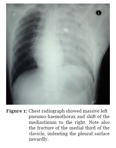
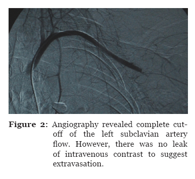
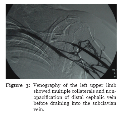
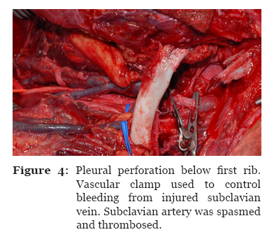
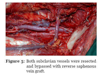
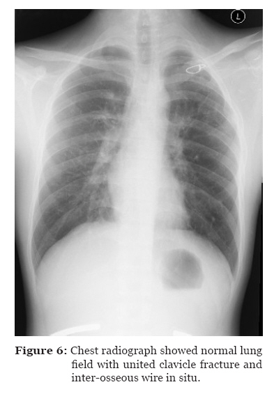
Malaysian Journal of Medical Sciences, Vol. 18, No. 2, 2011, pp. 74-77 Case Report Clavicle Fracture and Subclavian Vessels Disruption with Massive Haemothorax Mimic Intrathoracic Injury Wan Ismail Faisham1, Paiman Mohammad1, Haron Juhara2, Nik Mahdi Munirah2, Hassan Shamsulkamaruljan3, Ghazali Mohamad Ziyadi4 1 Department of Orthopaedic, School
of Medical Sciences, Universiti Sains Malaysia Health Campus, 16150 Kubang
Kerian, Kelantan, Malaysia Correspondence: Associate Professor Dr Faisham Wan Ismail MD (UKM), MMed Ortho (USM) Department of Orthopaedic School of Medical Sciences Universiti Sains Malaysia Health Campus 16150 Kubang Kerian Kelantan, Malaysia Tel: +609-767 6381 Fax: +609-766 4510 Email: faisham@kb.usm.my Submitted: 18 Apr 2010 Code Number: mj11026 Abstract We report a case of open fracture of the clavicle with subclavian artery and vein laceration and perforation of the parietal pleural below the first rib that caused massive haemothorax. Emergency thoracotomy and exploration followed by repair of both vessels were able to salvage the patient and the extremity. Keywords: bone fractures, clavicle, haemothorax, subclavian artery, trauma Introduction Fracture of the clavicle is common, accounting 5% of all fracture cases. Damage to the neurovascular structure associated with fracture of the clavicle, however, is rare and more frequently related to penetrating injuries (1). To the best of the authors’ knowledge, the incidence of open fracture of the clavicle and subclavian vascular injury with penetrating injury to the pleura has not been documented in the literature. Case Report A previously healthy 19-year-old helmeted man on a motorbike had a head-on collision with a lorry at the speed of approximately 100 km/h. He was brought by passers-by to our institution within an hour after the accident. The patient was alert and oriented, with continuous bleeding from a 4.0-cm wound in the upper left chest, which was packed with sterile gauze and compressive dressing. The left upper limb was dusky, with absence of radial and ulnar pulses. Emergency chest radiography revealed extensive left pneumo-haemothorax, with mediastinal shift to the right side and left medial third clavicle fracture with significant displacement (Figure 1). He also had left femur and segmental mandible fracture. A left tube thoracostomy initially returned 1000 mL of blood, with continuous drainage of 800 mL over 2-hour period. Aortic arch and selective left subclavian artery angiography revealed tapering of subclavian artery, with filling defect at the distal end due to spasm-associated complete thrombosis (Figure 2). Upper limb venography showed non-opacification of the distal cephalic vein and multiple collaterals seen from the cephalic and subclavian veins that resulted from compressive dressing (Figure 3). There was no evidence of extravasation of intravenous contrast from the vessels into the chest or around the injured area. While the patient was induced and under mechanical ventilation prior to the surgery, there was gushing of blood from the wound; local exploration at the time was highly suspicious of intra-thoracic bleeding. Thoracotomy through left posterolateral approach revealed a collection of 1500 ml of blood and clots, with a 2.0-cm laceration at the left middle lobe and apical lung contusion. There was 2.0 × 2.0 cm perforation of apical pleural below the first rib with avulsion fracture of the latter. Exploration of the subclavian vessels revealed 1.0-cm laceration of vein with active bleeding, segmental spasm of subclavian artery, and contused brachial plexus (Figure 4). Both artery and vein was resected and bypassed with reverse saphenous graft (Figure 5). Pleural perforation was repaired and medial clavicle was stabilised with inter-osseous wiring. The fractured femur and mandible were stabilised with plate and screwed 48 hours later. The patient was ventilated for 48 hours and tube thoracostomy was removed at day 3 post-operatively. There was presence of lower plexus neuropraxia, evidenced by weak handgrips, power of grade 3. Subsequent chest radiograph showed full lung expansion with minimal local pulmonary contusion. The patient returned home after 12 days of hospitalization with full strength and sensation of the left upper limb. At 3-month follow-up, his radial pulse was equal with no neurological deficits and the fractured clavicle had united with the help of the in situ wire (Figure 6). Discussion The initial approaches in subclavian vessels injury include aggressive resuscitation of hypovolaemic shock, assessment of other injuries, and diagnostic angiography, if time permits (2,3). External bleeding from the subclavian artery must be controlled rapidly, and associated pneumo-haemothorax must be managed by urgent chest thoracostomy. Direct pressure and compressive dressing are effective in controlling bleeding in urgent situation, particularly in the emergency department. Proper compression to the subclavian vessels injury produces tamponade, and this method is effective in most of the cases. Pleural perforation within the injured area can drain blood from the injured vessels to the pleural cavity, producing massive haemothorax; this condition may mislead the judgment that ongoing bleeding was controlled. This case highlighted that the mediastinum was a potential space with negative pressure that was able to collect huge amount of ongoing bleeding, which led to persistent hypovolaemic shock (3–6). Haemodynamically unstable patient with massive haemothorax should undergo immediate thoracotomy to obtain rapid vascular control (3–6). Angiography can be performed in more stable patients, and it is helpful in locating the injured area prior to surgical intervention (2). Massive haemothorax with intact aortic arch and proximal subclavian artery raised a question that injury can be caused by other thoracic vessels, which was difficult to diagnose and carries high mortality rate (3,4). The decision to perform a thoracotomy in this case was based on the findings of chest radiograph and blood gushing from thorax after ventilation as no option was left to control the bleeding. Furthermore, controlled haemostasis and definite identification of subclavian vessels injury was required for vessels reconstruction (1,4,6). Combination angiography and endovascular stent graft is also a viable option to minimized surgical morbidity with good outcome (5). The mediastinum and pleural space are potential areas to develop hematoma due to negative pressure. This will lead to false clinical judgment as external bleeding was well controlled despite of deteriorating haemodynamic status. Lung ventilation produces positive pressure and the haemothorax will be pushed through pleural perforation mimicking intrathoracic massive bleeding. Conclusion Clavicle fracture is known as a benign condition, but it can potentially lead to intrathoracic vascular injury and massive haemothorax. References
© Copyright 2011 - Malaysian Journal of Medical Science The following images related to this document are available:Photo images[mj11026f3.jpg] [mj11026f5.jpg] [mj11026f2.jpg] [mj11026f6.jpg] [mj11026f4.jpg] [mj11026f1.jpg] |
| |||||||||
