 |
| About Bioline | All Journals | Testimonials | Membership | News |
|
||||||
|
||||||
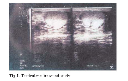
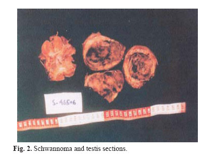
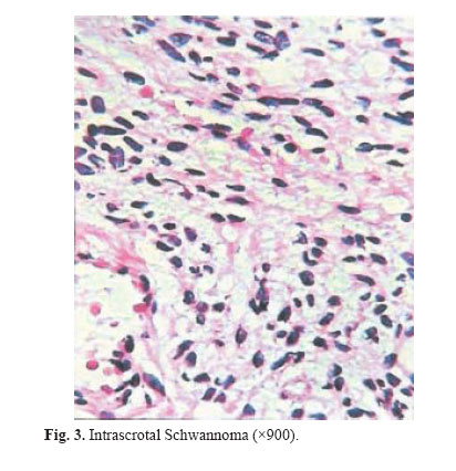
Medical Journal of the Islamic Republic of Iran , Vol. 18, No. 1, May, 2004, pp. 85-86 Case Reports HUGE PRIMARY INTRASCROTAL SCHWANNOMA: A CASE REPORT AND REVIEW OF THE LITERATURE ALI SHAMSA, M.D.,* AND ABBAS A. OMIDI, M.D.** From the Departments of *Urology and **Pathology, Ghaem Medical Center, Mashhad University of Medical Sciences, Mashhad, Iran. E-mail:shamsa@mums.ac.ir Code Number: mr04014 ABSTRACTTesticular schwannoma is a very rare benign scrotal tumor. It is a painless mass, but sometimes referred because of pain or sensory losses. Tumor markers are normal and radical orchidectomy is its best treatment. Here we present an unusual case of intrascrotal schwannoma in a 57 year old man, with a review of the literature. Keywords: Schwannoma, intrascrotal mass, neurinoma. INTRODUCTIONIntrascrotal masses are common in urologic practice. They consist of a wide spectrum of disease including infectious (epididymo-orchitis), vascular (varicocele), cystic (hydrocele) and solid (testicular tumors) masses. Treatment of these masses depends upon many factors, for example: nature of disease, definitive diagnosis, etc.We herein report a very rare and unusual case of intrascrotal non-testicular schwannoma. CASE REPORTA 57-year-old farmer with an 8-year history of painless right intrascrotal mass referred to us because of sudden severe pain in the mass. On physical examination, his right hemiscrotum had a large mass which did not transilluminate. His scrotal skin had a normal appearance, and he had no history of urinary infection. His ultrasound showed the right testis with a heterogenous solid mass attached very closely to the lower testis pole (Fig. 1). His blood AFP and βHCG were within normal limits. He was operated on the next day through an inguinal incision. The mass was fixed to the lower part of the testicle, so orchidectomy was carried out (Fig. 2). Macroscopic examination of the specimen revealed a small testis 1.5× 2 ×4 cm in diameter which was connected by fibroconnective tissue to an encapsulated mass with a diameter of 11× 8× 6.5 cm. The weight of the mass was 350 grams. Its surface was smooth; but its cut surface was lobulated and hemorrhagic with a greyish brown color. On microscopic examination, proliferation of spindle cells with neither atypia nor cellular anarchy (Antony A) was seen. On cross sections, the testis and epididymis were normal and intact, i.e. benign intrascrotal non-testicular schwannoma (Fig. 3). He was discharged from the hospital the next day and followed for 3 months with out any serious complication. DISCUSSIONSchwannomas are non- neural tumors which arise from Schwann cells. They are subdivided into benign and malignant (neurogenic sarcoma or malignant schwannoma) types.1 Benign schwannoma is also called neurilemmoma, neurinoma, schwanno-glioma, peripheral glioma, lemmocytoma and Schwann cell tumor.2 Benign schwannoma may occur anywhere in the central or peripheral nervous system. They may occur in any age and race but are common between 30-50 years of age without gender difference. Some of these tumors are associated with von Recklinghausen's disease; but most of them, like our case, are not.3 They are slow growing and occur most commonly on the flexor extremity, neck, mediastinum, thoracic cavity, retroperitoneum, posterior spinal roots and pelvis.1,4 Schwannomas are usually found incidentally, however, and motor and sensory losses are rare. Our case had no neurologic and/or urologic signs and symptoms except for a scrotal mass. The etiology of these tumors is unknown. It has a capsule, but, in our case, it was tightly adherent to the testis making it impossible to separate them. As far as our knowledge is concerned, this is the first case reported from Iran, and only the fifth case of intrascrotal non-testicular Schwannoma in the literature. But our case is different from the first (Aricola et al.4 ), second (Safak et al.3 ), third (Shimizu et al.5 ), and fourth (Amano et al.6 ) cases, because of a longer history of existing intrascrotal mass (8 years), size (11× 8 ×6.5 cm), weight (350 g), nature (benign) and location (close to testis). Malignant transformation of benign Schwannoma is possible, so orchidectomy and follow-up of the patient with periodic abdominal CT is advisable and highly recommended. REFERENCES
Copyright 2004 -Medical Journal of the Islamic Republic of Iran The following images related to this document are available:Photo images[mr04014f1.jpg] [mr04014f3.jpg] [mr04014f2.jpg] |
| |||||||||