search
for
 About Bioline  All Journals  Testimonials  Membership  News


Medical Journal of The Islamic Republic of Iran
National Research Centre of Medical Sciences of I.R. IRAN
ISSN: 1016-1430
Vol. 18, Num. 2, 2004, pp. 107-110

Medical Journal of the Islamic Republic of Iran , Vol. 18, No. 2, August, 2004, pp.107-110

CORRECTED QT DISPERSION IN CHILDREN WITH ISOLATED UNCOMPLICATED MITRAL VALVE PROLAPSE

ELAHEH MALAKAN RAD, AND NADER MOMTAZMANESH

From the Department of Pediatrics, Kashan University of Medical Sciences, Kashan, I.R. Iran.

Address: Kashan University of Medical Sciences, Ghotb Ravandi Blvd., Kashan 87154, I.R. Iran. Tel: (0361)-552999-8268752 Fax: (0361)-552999, malakan_rad@Kaums.ac.ir

Code Number: mr04019

ABSTRACT

Mitral valve prolapse (MVP) is a relatively frequent valvular abnormality. In both children and adults with MVP, an increased incidence of ventricular arrhythmias has been reported. QT dispersion, defined as the difference in duration between the longest QT interval and the shortest one, for a given set of electrocardiographic leads has been proposed as a sign of regional difference in cardiac repolarization and as a marker of increased risk of ventricular arrhythmias. This study was designed to compare the corrected QT dispersion (QTcD) in children with isolated uncomplicated prolapse with a normal age-and sex-matched group of children. Twenty children with MVP (mean+SD age: 8+5 years) and thirty normal children enrolled in the study. Corrected QT dispersion was manually calculated on their surface electrocardiogram in all of them. QTcD was 39+14.7 and 40+15.2 milliseconds respectively. An unpaired Student t-test was performed. There was no significant difference between! the mean value of the two groups (p value<0.01). The findings of our study, besides the reports of increased QT dispersion in adults, suggest that increased QT dispersion in patients with MVP is a time-evolved phenomenon and does not exist from the very early years of age.

Keywords: Corrected QT dispersion, Mitral valve prolapse, Children.

INTRODUCTION

Ventricular arrhythmias are seen in patients with mitral valve prolapse (MVP) with increased frequency.1-2 The contribution of this heterogeneity of the repolarization process within the myocardium for triggering ventricular arrhythmias has also been already recognized.3,4,5 The existence of an abnormal pattern of ventricular repolarization has been exclusively demonstrated.6-7

Corrected QT dispersion (QTcD), measured in the 12- lead surface electrocardiogram is a simple approach to the evaluation of ventricular repolarization.8 Despite relatively extensive research on QT dispersion in adults with mitral valve prolapse,9-15 to our knowledge no or perhaps few studies have addressed the issue of QT dispersion in children with MVP. The aim of this study is to determine whether QT dispersion of children with MVP is different from normal controls.

MATERIAL AND METHODS

A resting 12-lead electrocardiogram was obtained from 20 children with uncomplicated isolated MVP, including 12 females and 8 males (aged 3 to 14 years,mean [±SD]age:8±5 years). Only children who had both the echocardiographic and auscultatory findings of MVP were included.On echocardiography, mitral valve prolapse was diagnosed only when there was definite prolapse of the leaflets observed in at least two echocardiographic planes (parasternal long-axis and four chamber views).16 None of our patients had MR, either on auscultation or echocardiography. Prolapse was defined as displacement of one or both mitral leaflets by more than 2 millimeters above the high points of the mitral annulus. Individuals who were on drug therapy that could potentially affect QT characteristics were excluded. Thirty age- and sex-matched healthy children were enrolled in the study as control. Children who were referred for the evaluation of a cardiac murmur and following thorough echocardiographic study their murmur was proved to be innocent were assigned as the control group. Corrected QT dispersion (QTcD) was measured in both groups (MVP and control). A three channel electrocardiographic recorder (Esaote, model P80, manufactured by SAG, made in Switzerland and distributed by Esaote S.P.A., Italy) was used. ECGs were recorded at a speed of 25mm/s and amplitude of 10mm/mv. The QT and preceding PR intervals of three consecutive sinus beats were measured in a range of nine to twelve leads17-19 The leads with flat ST-T waves were excluded from analysis.20 The QT and RR intervals were measured manually with calipers! by a single observer. Thereafter repeatability of the QTcD measurements was tested by a second observer. QT interval was taken from the onset of the QRS to the end of T wave (i.e., return to the T/P baseline). If U waves were present, the QT interval was measured to the nadir of the curve between the T and U wave. The QTcD was calculated by the method of Bazett21 (QTc= QT

√RR). Dispersion of the QTc(QTcD)was defined as the difference between the maximum and minimum QTc interval occuring in any of the 12 leads. QTcD is expressed as milliseconds.

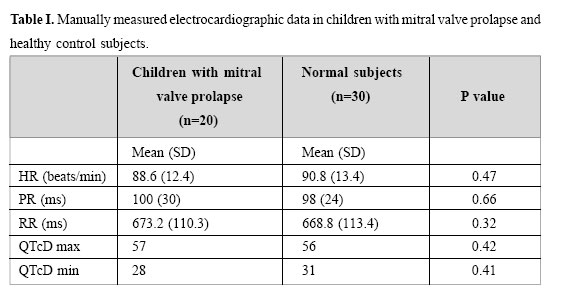
Data are expressed as mean± SD. Unpaired one-tailed Student’s t test was used for testing the difference between the two groups. A P value<0.05 was considered significant.

RESULTS

None of our patients had ventricular arrhythmias on surface electrocardiogram. QTcD was 39±14.7 and 40±15.2 milliseconds in the two groups of control and children with MVP respectively. Mean values of the two groups were compared using unpaired Student’s t-test. The QTcD was not significantly different in children with MVP. Other ECG findings are mentioned in Table I.

DISCUSSION

This study demonstrates that in children with uncomplicated isolated MVP, increased QTcD is not as frequent as in adults. Indeed none of our patients showed a significantly different QTcD in comparison with our normal children group.

Ventricular arrhythmias are common in adults with MVP. PVCs and complex PVCs has been reported to occur in 58% to 80% and approximately 50% of adults with MVP respectively.8 Leclercq et al. reported ventricular arrhythmias in 70% of their patients.22 Various causes have been cited for the occurrence of ventricular arrhythmias, including autonomic dysfunction, papillary muscle traction, abnormal QT dispersion, mechanical stimulation of myocardium by leaflets, abnormal innervation of floppy mitral valve and so on.23 Babuty et al. evaluated 58 adult patients with MVP and concluded that the main determinant factors of complex ventricular arrhythmias in these patients are patient age and mitral regurgitation.15 Tieleman et al. studied 32 patients with echocardiographically documented mitral valve prolapse and showed that QT dispersion on the 12 lead surface electrocardiogram was greater in patients with MVP with ventricular arrhythmias than in normal controls.10

To our knowledge, there is no study that has specifically addressed the issue of QT dispersion in children with uncomplicated isolated MVP. Wroblewska – Kaluzewska et al. analyzed arrhythmia and ST –T abnormalities in children with mitral valve prolapse. Only one

age
patient out of their 78 cases had QTc interval prolongation. They did not evaluate the QTc dispersion.24 Although several studies in adult population have shown increased QT dispersion in patients with MVP and its contribution to ventricular arrhythmias.9-15 Unfortunately there is no data about the QT dispersion of these patients at their childhood ages.Although in normal individuals QT dispersion does not vary significantly with 25-26 our results raise the question whether abnormal QT dispersion in patients with MVP is a phenomenon that evolves over relatively long periods of time, i.e. passage from childhood to adulthood. In order to test the truth of such a hypothesis, it seems that serial measurement of QTcD at time intervals of several years can be helpful. Overall, age appears to be an important factor in the development of complications of MVP27-29 Bisset et al. found no progression of mitral regurgitation and no sudden death among 119 children monitored for a mean age of seven years starting at mean age of 10 years30 Although the mechanism or mechanisms of abnormal QT dispersion in patients with MVP have not been specifically and clearly delineated, but in general autonomic dysfunction29 or ischemia32 can play an important role. Since most complications of mitral valve prolapse develop over time and become manifest in adulthood, it is likely that the normal QT dispersion of childhood is also one of the several benign features of the disease that is subject to change with passage of time.

Another explanation might be sought in the real importance and clinical potential of QTcD. Malik et al. reported that although QT dispersion is increased in cardiac patients compared with healthy subjects and the prognostic value of QT dispersion has been reported, values are largely overlapping, both between healthy subjects and cardiac patients and between patients with and without adverse outcome.They added that QT dispersion is a crude and approximate merasure of abnormality of the complete course of repolarization.33

In conclusion, we found that QTcD in children with MVP does not differ significantly from normal controls. This may explain the low incidence of ventricular arrhythmias in this age period. Considering several reports of increased QT dispersion in patients with MVP,it seems that the abnormality of QT dispersion is a phenomenon that occurs over several years and is not a congenital electrophysiologic abnormality.

REFERENCES
  1. Bobkowski W, Siwinski A, Gorzna H, Niedbalski R, Paluszak W: Dysrhythmias documented by 48-hour electrocardio-graphic monitoring in children wwith mitral valve prolapse. Pediatr Pol 71(6): 493-7, June 1996.
  2. Nowak A, Czerwionka-Szaflarska M: Arrhythmias in children with mitral valve prolapse syndrome. Pediatr Pol 71(6): 499-504, June 1996.
  3. Glancy JM, Garrat CJ, Woods KL, et al: QT dispersion and mortality after myocardial infarction. Lancet 354: 945948, 1995.
  4. Grimm W, Steder U, Menz V, et al: Clinical significance of increased QT dispersion in the standard 12-lead ECG for arrhythmia prediction in dilated cardiomyopathy. Pacing Clini Electrophysiol 19(11): 1886-1889, 1996.
  5. Day CP, McComb JM, Campbell RWF: QT dispersion: an indication of arrhythmia risk in patients with long QT intervals. Br Heart J 63: 342-4, 1990.
  6. De Ambroggi L, Bertoni T, Locati E, et al: Mapping of body surface potentials in patients with idiopathic long QT syndrome. Circulation 74: 1334-1345, 1998.
  7. De Ambroggi L, Negroni MS, Monza E, et al: Dispersion of ventricular repolarization in the long QT syndrome. Am J Cardiol 68: 614-620, 1991.
  8. Schwartz Peter J, Priori SG., Napolitano C: The long QT syndrome. In: Zipes Douglas P, Jalife J, (editors), Cardiac electrophysiology, from cell to bedside. Philadelphia: W.B. Saunders, pp. 563-568, 2000.
  9. Kulan K, Komsuoglu B, Tuncer C, Kulan C: Significance ofQT dispersion on ventricular arrhythmias in mitral valve prolapse. Int J Cardiol 54(3): 251-7, June 1996.
  10. Tieleman RG, Crijns HJ, Wiesfeld AC, Posma J, Hamer HP, et al: Increased dispersion of refractoriness in the absence of QT prolongation in patients with mitral valve prolapse and ventricular arrhythmias. Br Heart J 73(1): 37-40, June 1995.
  11. Ulgen MS, Biyik I, Karadede A, Temamogullari AV, Alan S, Topra KN: Relation between QT dispersion and ventricular arrhythmias in uncomplicated isolated mitral valve prolapse. Jpn Circ J 63(12): 929-33, Dec 1999.
  12. Maraglino G, Sturaro M, Toniolo G, Accurso V, Pastore G, Palatini P: Relation between ventricular late potentials and ventricular arrhythmias in mitral valve prolapse. G Ital Cardiol 24(8): 957-64, 1994.
  13. Corrodo D, Basso C, Nava A, Rossi L, Thiene G: Sudden death in young people with apparently isolated mitral valve prolapse. G Ital Cardiol 27(11): 1097-105, Nov 1997.
  14. Zouridakis EG, Parthenakis FI, Kochiadakis GE, Kanoupakis EM, Vardas PE: QT dispersion in patients with mitral valve prolapse is related to the echocardiographic degree of the prolapse and mitral leaflet thickness. Europace 3(40: 292-8, Oct 2001.
  15. Babuty D, Cosnay P, Breuillac JC, Charniot JC, Delhomme C, Fauchier L, Fauchier JP: Ventricular arrhythmia factors in mitral valve prolapse. Pacing Clin Electrophysiol 17(6): 1090-9, Jun 1994.
  16. Snider A, Rebecca Serwer GA, Ritter Samuel B: Echocardiography in pediatric heart disease. St. Louis: Mosby Year Book, Second edition, pp. 398-399, 1998.
  17. Moss AJ, Schwartz PJ, Crampton RS, Locati E: Carleen E: The long QT syndrome:a prospective international study. Circulation 71: 17-21, 1985.
  18. Suys BE, Huybrechts SJA, Wolf D, Beeck L, Matthys D, Overmeire V, et al: QTc interval prolongation and QTc dispersion in children and adolescents with type 1 diabetes. J Pediatr 141: 59-6, 2002.
  19. Barr CS, Naas A, Fenwick M, Struthers AD: Enalapril reduces QTc dispersion in mild congestive heart failure secondary to coronary artery disease. Am J Cardiol 79: 328333, 1997.
  20. Kors JA, Herpen GV: Measurement error as a source of QT dispersion: a computerized analysis. Heart 80: 453458, 1998.
  21. Park MK, Guntheroth WG, (editors): How to read pediatric ECGs. St Louis: Mosby Year Book, p. 54, 1992.
  22. Leclercq JF, Malergue MC, Milosevic D, Rosengarten MD, Attuel P, Coumel P: Ventricular arrhythmias and mitral valve prolapse. A study of 35 cases. Arch Mal Coeur Vaiss 73(30): 276-87, 1980.
  23. Boudoulas H, Wooley C: The floppy mitral valve, mitral valve prolapse, and mitral valvular regurgitation. In: Allen H, Clark E, Gutgesell H, Driscoll D, (editors), Moss and Adam’s heart disease in infants, children, and adolescents. Philadelphia: Williams&Wilkins, pp. 947-969, 2001.
  24. Wroblewska-Kaluzewska M, Piorecka-Makula A, Tomik A: Arrhythmia and repolarization in children with mitral valve prolapse. Wiad Lek 53(9-10): 513-7, 2000.
  25. Vialle E, Albalkhi R, Zimmerman M, Friedli B: Normal values of signal-averaged electrocardiographic parameters and QT dispersion in infants and children. Cardiol Young 9(6): 556-61 Nov 1999.
  26. MacFarlane PW, McLaughlin SC, Devine B, Yang TF: Effect of age, sex, and race on ECG interval measurement. J Electrocardiol 27 Suppl: 9-14, 1994.
  27. Smallhorn J, Macartney FJ: Mitral valve anomaliesand supravalvar mitral ring. In: Anderson RH, Baker EJ, Macartney FJ, Rigby ML, (editors). Paediatric Cardiology. London: Churchill Livingstone, pp. 11531166, 2002.
  28. Alexander ME: Ventricular arrhythmias in children and young adults. In: Walsh EP, Saul JP, Triedman JK, (editors), Cardiac arrhythmias in children and young adults with congenital heart disease. Philadelphia: Williams& Wilkins, pp. 224-234, 2001.
  29. Kanter RJ: Mitral valve prolapse, In: Gillette PC, GarsonA, (editors), Clinical Pediatric Arrhythmias. Philadelphia: W.B. Saunders Company, pp. 263-264, 1999.
  30. Bisset GS, Schwartz DC, Meyer RA, et al: Clinical spec-trum and long-term follow-up of isolated mitral valve prolapse in 119 children. Circulation 62: 423-429, 1980.
  31. Haapalahti P, Makijarvi M, Montonen J, Korhonen P, Salorinne Y, Oikarinen L, Vitasalo M, Toivonen L: Effects of cardiovascular autonomic function tests on QT dispersion in the 12-lead electrocardiogram of healthy patients. Electrocardiol 33(4): 321-7, Oct 2000.
  32. Roukema G, Singh JP, Meijs M, Carvalho C, Hart G: Effect of exercise-induced ischemia on QT interval dispersion. Am Heart J 135: 88-92, 1998.
  33. Malik M, Batchvarov VN: Measurement, interpretationand clinical potential of QT dispersion. J Am Coll Cardiol 15; 36(6): 1749-66, Nov 2000.

Copyright 2004 -Medical Journal of the Islamic Republic of Iran


The following images related to this document are available:

Photo images

[mr04019t1.jpg]
Home Faq Resources Email Bioline
© Bioline International, 1989 - 2024, Site last up-dated on 01-Sep-2022.
Site created and maintained by the Reference Center on Environmental Information, CRIA, Brazil
System hosted by the Google Cloud Platform, GCP, Brazil