 |
| About Bioline | All Journals | Testimonials | Membership | News |
|
||||||
|
||||||
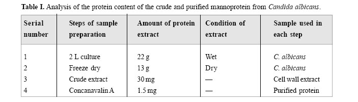
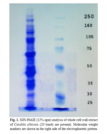
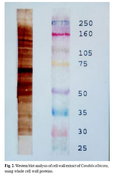
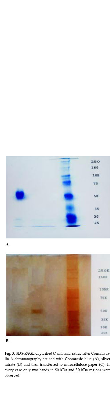
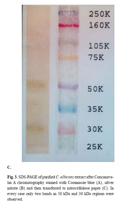
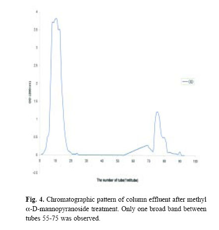
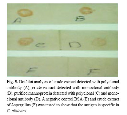
Medical Journal of the Islamic Republic of Iran , Vol. 18, No. 2, August, 2004, pp. 167-172 PURIFICATION AND CHARACTERIZATION OF CELL WALL MANNOPROTEINS OF CANDIDAALBICANS USING INTACT CELL METHOD Z. FARAHNEJAD,* M.J. RASAEE,** H. YADEGARI,* AND M. FROUZANDEH MOGHADAM*** From the *Department of Medical Mycology, the **Department of Clinical Biochemistry, and the ***Department of Medical Biotechnology, School of Medical Sciences, Tarbiat Modarres University, Tehran, I.R. Iran. Address of Correspondence: Mohammad Javad Rasaee, Tel.: +98-21-8013030; Fax: +98-21-8006544. E-mail: rasaee_m@modares.ac.ir. Code Number: mr04031 ABSTRACTVirulence of the opportunistic yeast, Candida albicans , involves the interplay of many complex changes including the yeast-hyphae transition, which mainly involves protein changes. Cell wall mannoproteins are found to be the main cause of adherence of C. albicans to epithelial cells in the first step of an infection process. In the present study, cell wall mannoproteins of intact yeast were purified using a simple treatment of yeast with mercaptoethanol and sodium dodecyl sulfate followed by Concanavalin A chromatography. Both electrophoretic analysis of the column effluent and Western blot analysis using polyclonal and monoclonal antibodies showed the presence of mannoproteins with molecular weight in the range of 30-50 kDa. Dot blot analysis of the purified antigen with the polyclonal and monoclonal antibodies prepared in this study showed that outer membrane mannoprotein antigens were obtained successfully following the above simple purification strategy. Keywords: Candida albicans , Mannoproteins, Purification, Characterization. INTRODUCTIONCandida albicans is a dimorphic opportunistic pathogen that grows as a yeast or as a mycelial fungus depending on the environmental conditions.1 The major components (80-90%) of cell wall of C. albicans are carbohydrates, mannose or polymers of mannose covalently associated with proteins to form glycoproteins, also referred to as manno-proteins. These proteins contain β-glucans that are branched polymers of glucose containing β-1,3 and β-1,6 linkages and chitin, which is an unbranched homopolymer of N-acetyl-D-glucoseamine containing β-1,4 bonds. Proteins (6-25%) and lipids (1-7%) are also present as minor wall components.2,3 β-Glucans and chitin (0.6-9%) are also present as structural components of the wall (47-60% by weight). Mannoproteins are considered as the most important antigenic component of Candida strains composing 10-30% of the cell wall.1 This group of proteins are mainly composed of carbohydrate polymannose containing more than 150 strongly bonded mannosyl units. Mannoproteins play an important role in the process of adherence of Candida strains to mucosal surfaces which allows the organism to cause infection. Therefore the study of the cell wall proteins of C. albicans is of immense importance in order to understand the actual mechanism of infection leading to probable prevention or treatment of the disease. In several studies, cell wall polymers have been extracted under controlled degradation of whole cells or of isolated walls using various enzymes for digestion.4-6 As an alternative method, solubilization of cell wall materials using chemicals such as mercaptoethanol (ME), dithiothreitol (DTT), and ethylene diamine has also been reported.7-9 Analysis of antigen expression with polyclonal and monoclonal antibodies has revealed the complex antigenic composition of the surface of C. albicans cells. These studies suggested that mannoproteins are the main antigenic cell wall components,10 which may subsequently be used as basic antigen during antibody development. In this case, it is obvious that natural conformation of dominant antigens such as surface antigens is of extreme importance. In this study, we have attempted to purify the surface mannoprotein of C. albicans using intact cells with chemical treatments (sodium dodecyl sulfate (SDS) and ME) followed by Concanavalin A affinity chromatography. In this way, the major conformational characters of mannoprotein were left unaltered and hence the mannoproteins were purified in their natural conformation. MATERIAL AND METHODS Organism isolationCandida albicans was isolated, cultured, and maintained from patients with vaginitis. The isolated strain was identified by using Candida check (Iatron laboratories, Tokyo, Japan) with an additional germ tube test and by examining morphological characteristics. Microbiological observations of pseudohyphae, hyphae and chlamydospores were made on cornmeal Tween 80 agar incubated at 35°C for 3 days. Culture medium GYEP containing 2% glucose, 0.3% yeast extract and 0.1% peptone (supplemented with penicillin 100 IU/mL and streptomycin 100 µg/mL) were used for C. albicans. Glucose and peptone solutions were autoclaved (Solution A) while water-dissolved yeast extract was filtered through 0.2 µm filters (Solution B). Solutions A and B were mixed and reconstituted to 2 L. The resulting solution was divided into 4 Erlenmeyers and 2 mL of C. albicans suspension was added to each container. The containers were incubated and shaken (100 rpm) at 29°C for 48 h. At the end of incubation time, samples were centrifuged (×800 g, 5°C), the pellet was collected, washed three times with D.D. water, centrifuged three days after each washing step (×800 g, 5°C), and finally it was stored at -20°C. Out of 2 L of culture medium about 22 g of C. albicans was obtained. Antiserum preparationCandida albicans serotype A from vaginitis cultures was used. These isolates were grown on Sabouraud’s dextrose agar plates, washed using saline, harvested by centrifugation and washed three times in sterile saline. Balb/c mice were inoculated subcutaneously with a 1:1 emulsion of 106 cell suspension in saline and complete Freund’s adjuvant. Two weeks later the mice received another inoculation IP with a 1:1 emulsion of 106 cell suspensions in saline and incomplete Freund’s adjuvant. Booster injections were given every 15 days for two months. The mice were checked for antibody production and titer after each bleeding episode which followed after each injection by ELISA method. ELISA for detection and titration of antibodyAn ELISA method was developed and used for detection of antibody. In this method, the crude extract, purified mannoprotein and irrelevant antigens (such as BSA) were coated onto the wells of a microtiter plate in similar concentrations. Wells were washed (PBS, 10 mM, containing 0.05 Tween 80), blocked with PBS containing 0.3% gelatin and added with dilutions of antibody in duplicates (1:500, 1:1000, 1:2000) and incubated for 2 h. A dilution of normal mouse serum, 1:300, was used to indicate nonspecific binding. At the end of incubation time, wells were emptied, washed four times and added with a 1:2000 dilution of anti-mouse immunoglobulin labeled with HRP (Sigma Chemical Co., St. Louis, MO, USA), incubated for 1 h, and washed with Tween20 containing PBS buffer (0.05%). Finally tetramethyl benzedene substrate solution was added and the optical density was detected at 450 nm. Preparation of cell wall extractsThis was performed according to the method of Casson et al.11 with minor modifications as follows. C. albicans suspension was treated with lysis buffer (containing 2% SDS and 5% ME dissolved in water). The resulting suspension was vortexed vigorously, incubated in boiling water for 5 min and cooled in ice bath for 2 min. The resulting solution was centrifuged at 14,000 g for 10 min, the supernatant was removed and measured for protein content using Bradford protein assay procedure. The resulting solution was dialyzed against 0.1 M acetate buffer solution, pH=6.0 and the clear solution was freeze dried and finally electrophoresed on polyacrylamide gel. Affinity chromatographyIn order to further purify the crude extract, a Sepharose 4B Concanavalin A packing material was used. Ten milliliters of Concanavalin A beads was prepared (as the instruction procedure provided by the manufacturer, Sigma Chemical Co.), and were packed in a 10×1.5 cm column, washed and equilibrated with acetate buffer (0.1 M, pH=6 containing sodium chloride 1 M, calcium chloride, manganese chloride and magnesium chloride, 1 mM). The crude extract (400 mg) was dissolved in a minimum quantity of equilibration buffer (1 mL), loaded onto the column and washed extensively with acetate buffer (0.1 M, pH=6) until no protein was detected in the column effluent. Mannoproteins were then eluted using acetate buffer containing methyl α-D-mannopyranoside (0.3 M) with a slow rate of 15 mL/h. Samples were collected in 2 mL increments and were analysed by SDS polyacrylamide gel electrophoresis (SDS-PAGE) followed by Western blotting analysis using polyclonal antibody prepared against cell wall proteins. Further characterization of mannoprotein prepared in the above procedures was performed by dot blot analysis. RESULTSTable I indicates the amount of crude and purified mannoprotein obtained in different steps. It was found that out of 2 L of liquid culture containing 22 g of wet yeast, 1.5 mg of mannoprotein could be obtained. Figure 1 shows the result of SDS-PAGE of the crude extract (after SDS and 2 ME treatment), where around 20 bands (from 25 kDa up to 250 kDa) were obtained. When these extracts were blotted and detected with polyclonal antibodies, 19 bands (from 30 kDa to 250 kDa) were observed (Figure 2). Figurea 3a and 3b are SDS-PAGE results for crude extract being further purified using Concanavalin A column. Here the result indicated that only two bands (50 kDa and 30 kDa) both in Coomassie blue staining (Figure 3a) and silver nitrate staining (Figure 3b) were presented. When these proteins were transferred to nitrocellulose paper and stained with polyclonal antibody, the same two bands (a strong 50 kDa and a relatively weak band at 30 kDa) were observed (Figure 3c). Figure 4 shows the chromatographic pattern of Concanavalin A purification of mannoprotein. Here it was found that only one peak containing mannoprotein (a 50 kDa and a 30 kDa protein as was shown in electrophoresis) was eluted after methyl α-D-mannopyranoside (0.3 M) treatment of the column between tubes 55 to 75. The results of dot blot analysis are shown in Figure 5. In these results it was indicated that the crude extract reacted with monoclonal antibodies of factor 6 (specific for C. albicans obtained from Iatron laboratories, Tokyo, Japan) and polyclonal antibodies prepared in this study (Figures 5a and b). Further, it was shown that purified antigen (mannoprotein) reacted with the same combination of antibodies mentioned as above in a comparably lesser extent (Figures 5c and d) when an equal amount of antigen was blotted. However no reaction was detected between the antibodies when BSA or crude extract of Aspergillus were used as antigen. These results indicated that the antigens prepared in this study only reacted with monoclonal and polyclonal antibodies prepared for C. albicans. DISCUSSIONWe have shown that the extraction of mannoprotein from the intact cell wall of yeast using chemical method and affinity chromatography with Concanavalin A has more advantages than enzyme digestion. Using this procedure, we expected that purified antigens should retain main epitope features and conformational characters such that they may be successfully used as immunogen and antigen base for assay development. The simple and gentle treatment of C. albicans with SDS and 2 ME released a varied array of material from the cell wall. Many reports on analysis of cell wall extracts of Candida albicans are available. Ponton and Jones (1986) used DTT, DTT with protease, β-glucuronidase and chitinase to release the wall components.8 Casanova et al. (1989) used SDS followed by zymolyase treatment.12 Yadegari et al. (2001) reported the use of SDS followed by Concanavalin A and DEAE ion exchange chro-matography.13 Casanova and Chaffin (1991) examined different methods for cell wall release of glycoproteins of Candida albicans .11 In their experiments they used 2 ME, zymolyase, SDS, boiling and a combination of these reagents and physical conditions. In all these procedures an array of proteins detected by SDS-PAGE starting from 20 kDa up to 400 kDa have been reported.14 After performing Concanavalin A affinity chromatography we found just two bands in the region of 55 kDa (a predominant band) and 30 kDa (a weak band). This showed that mannoprotein may be purified in a simple procedure as explained. Western blot analysis confirmed this finding and the antibodies prepared against whole yeast extract, although reacting with 21 bands in crude extract, reacted with 2 bands after the purification procedure was carried out. Using monoclonal antibody prepared against epitope 6 with purified antigen showed that the antigens were reactive towards this antibody in dot blot analysis. We therefore concluded that mannoproteins of low molecular weight (30-55 kDa) may be purified by a two-step treatment explained in this work. REFERENCES
Copyright 2004 -Medical Journal of the Islamic Republic of Iran The following images related to this document are available:Photo images[mr04031f3.jpg] [mr04031f2.jpg] [mr04031f3c.jpg] [mr04031f1.jpg] [mr04031f3a.jpg] [mr04031f4.jpg] [mr04031f5.jpg] [mr04031f3b.jpg] [mr04031t1.jpg] |
| |||||||||