 |
| About Bioline | All Journals | Testimonials | Membership | News |
|
||||||
|
||||||
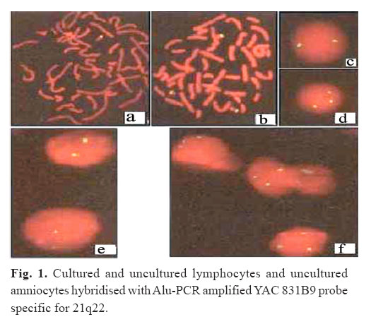
Medical Journal of the Islamic Republic of Iran , Vol. 19, No. 2, August, 2005, pp. 165-168 Basic Science in Medicine A PROSPECTIVE STUDY ON RAPID PRENATAL DIAGNOSIS OF TRISOMY 21 IN UNCULTURED AMNIOCYTES USING INTERPHASE FLUORESCENCE IN SITU HYBRIDISATION AND AN ALU–PCR AMPLIFIED YAC CLONE S.M. MOHADDES, Ph.D.1,2,3 S.M. TABATABAEI, M.Sc.,3 AND J. MOHSENI, M.Sc.3 From the 1Medical Genetics Unit, Faculty of Medicine, Tabriz University of Medical Sciences, Tabriz, the 2Islamic Azad University of Tabriz, Faculty of Medical Sciences, Tabriz, and the 3Medical Genetics Lab, Academic Center for Education, Culture and Research, Tabriz, Iran. Address for correspondence: S.M. Mohaddes Ardebili, Medical Genetic Unit, Department of Biochemistry, Faculty of Medicine, Tabriz University of Medical Sciences, Golgasht Ave, Tabriz-Iran. Tel/Fax: 0411-3344274, e-mail: 1) mohaddes@tbzmed.ac.ir 2) mohaddesmo@yahoo.com Code Number: mr05012 ABSTRACT Background: Herein we report the result of a prospective study directly comparing aneuploidy detection of chromosome 21 by fluorescence in situ hybridisation in interphase nuclei with the results obtained by cytogenetic analysis. Keywords: Down syndrome, Interphase FISH, Alu-PCR, YAC Clone. INTRODUCTION Down syndrome, the leading cause of genetically determined mental retardation, is amongst the most common genetic defects, occurring in approximately 1 in 700 births.1 The incidence of the disease increases exponentially in women over 35 years of age. Prior to birth, Down syndrome and other aneuploidies can be detected by amniocentesis and karyotyping.2 Despite the classical cytogenetic banding techniques having been extremely valuable in the assessment of chromosome number and structure, they have suffered from several limitations. First, detailed chromosome banding analysis can only be performed using high quality chromosome spreads, which are not always available. Secondly, for prenatal diagnostic applications, diagnosis is labor-intensive and time-consuming as it depends on the culture of fetal cells and the analysis of metaphase chromosomes.3,4 It has been shown that fluorescence in situ hybridisation can detect the number of copies of a particular chromosome present in interphase nuclei.5,11 The major advantage of this technique is that there is no requirement for cell culture and hence the results can be available in two days. The technique has important applications for the aneuploidy analysis of fetal chromosome abnormalities if it can be shown to be reliable in uncultured amniotic fluid cells. However chromosome 21 analysis in interphase appears to be more complicated than for many other chromosomes, as there is no reliable chromosome 21-specific repeat probe available. To overcome these problems the Alu-PCR product of a chromosome 21-specific YAC were used in this study to prenatally detect the number of chromosome 21 copies on uncultured amniocytes by FISH. The results obtained from application of the technique on 214 uncultured amniotic fluid samples revealed high detection efficiency on cell preparations. MATERIAL AND METHODS Sample preparationAbout 17-20 mL of amniotic fluid samples was received through gynecology hospitals for each patient who was at increased risk of a Down syndrome conception. About 15 mL of each sample was assigned by a lab code number and used for amniocyte culture according to standard cytogenetic techniques. The remaining 2-5 mL of each sample was detected by a different identification number and used for uncultured amniocyte preparation as described by Klinger et al.12 Uncultured amniocytes in PBS were dispensed on to 3-aminopropyl triethoxy silane-coated slides at 37°C (35µL vol/slide), two volumes of d. H20 pre-warmed at 37°C were added and incubated at 37°C for 15 min. The hypotonic solution was carefully decanted and replaced by 100 µL of 30% 3:1 fix (methanol: acetic acid) and 70%, 75mM KCl for 5 min at room temperature. This solution was carefully decanted and fresh 3:1 fix was dropped on to the slide from a height of 60 cm. Excess fix was decanted and slides dried for 5 min at 60oC, dehydrated through alcohol series (50%, 70%, 90% and 100%), air dried and stored at -20oC until required. Probe preparationTwo Alu primers: BK-33 (5'CTGGGATTACAGGCGTGAGC-3') priming to the 5' end of the Alu consensus sequences (nt positions 15-34) and SR1 (5'-CCACTGCACTCCAGCCTGGGG-3') close to the 3' end (nt position 241-261)13 were used to selectively amplify the chromosome 21 specific DNA sequence inside of the YAC: 831B9. The PCR assay was performed as described by Lengauer et al.14 with small modifications. 100 ng of the primer were each at a concentration of 0.25 µM in a total volume of 50 µL PCR buffer containing 250 µM of each of the four dNTPs, and 2.5 units of Taq polymerase (perkin-Elmer/cetus). After an initial denaturation at 96°C for 5 min, 30 cycles of PCR were carried out with denaturation at 96°C for 1 min, annealing at 37°C for 30s and extension at 72°C for 6 min. A 10 min extension was performed at the end of the last cycle. Ten-microlitre aliquots of amplified DNA sequences were fractionated by electrophoresis in 1.3% agarose gel in 1x T.B.E. (0.9 M Tris-HCl, 0.9 M boric acid and 20 mM EDTA). PCR products were ethanol precipitated, dissolved in TE (10 mM tris-HCl, 1mM EDTA, pH 8), and used for nick translation with biotin-11-dUTP. The labelled DNA was used as a probe for FISH. Chromosome in situ suppression hybridisationChromosomal in situ suppression (CISS) hybridization and probe detection with fluorescein isothiocyanate (FITC) conjugated to avidin were carried out according to Carter et al.15 with the following modifications: For hybridization 100-150 ng of Alu-PCR amplified YAC DNA was used as probe after pre-annealing with 100 ng of human placental DNA. The signals were amplified once. Cells were counter stained with 0.4 µg/mL 4,6 diamino-2phenylindol-dihydrochloride (DAPI) and 0.2 µg/mL propidium iodide in mounting medium AF1 (Citiflour Ltd) and were evaluated with conventional fluorescence microscope. RESULTSThe hybridisation and detection conditions were optimized using cultured lymphocytes. Various concentrations of probe and competitor DNA were investigated to achieve intense signals specific for chromosome 21 with little background. Figure 1a demonstrates a cultured lymphocyte from a normal individual and Figure 1b a cell from an individual with trisomy 21 hybridised with probe 831B9. In all experiments strong signals were observed on both chromatids of chromosome 21 at the expected locus on the long arm (21q22). To evaluate the detection efficiency of approach, the probe was initially hybridised to an unselected series of twenty uncultured lymphocytes and the results were rechecked by lymphocyte culture and GTG-banding for each sample. Eighteen samples were correctly scored as normal displaying two distinct signals specific for chromosome 21 on an average of 94 per cent of the hybridised cells (Figure 1c). Two samples showed three signals on an average of 87 per cent of hybridised cells and were correctly identified as trisomy 21 (Figure 1d). Figure 3.11a and b diagrammatically illustrates the detection efficiency of probe 831B9 on uncultured normal and abnormal lymphocytes respectively. The optimised procedure was applied to uncultured amniocytes, to detect the copy number of chromosome 21 in interphase nuclei. A total of 214 amniotic fluid samples were analysed in a blind fashion. The hybridisation signals were analysed using a conventional epifluorescence microscope and the results were compared to those obtained by traditional cytogenetic assay for each sample. One-hundred and ninety-nine samples showed two distinct signals on an average of 90.5 per cent of randomly evaluated nuclei and correctly detected as normal when compared to the results obtained from GTG-banding assay. Seven samples were revealed to be trisomic for chromosome 21 with a detection efficiency of 87 percent and confirmed by cytogenetic analysis (Figure 1e). One of the samples was shown to be normal using interphase FISH, however the cytogenetic assay revealed a Robertsonian translocation between long arms of chromosomes 14 and 21. The remaining 7 samples failed to produce a result owing to poor quality of the preparation and maternal cell contamination (Figure 1f). No false positive results was obtained in this study. DISCUSSIONThe most common chromosomal abnormality in newborns is Trisomy 21, with an incidence of 1/700. Prenatal diagnosis is routinely offered to women at increased risk of having a child with chromosomal abnormality, the most common indications being advanced maternal age or positive screening results based on biochemical marker screening and ultrasound evaluation.16,17 Conventional cytogenetic techniques based on banding of metaphase chromosomes are accurate and can often detect subtle rearrangements. However the time required to perform an analysis is around 2 weeks under the best circumstances. Methods that allow rapid and accurate detection of the major fetal aneuploidies are valuable, since they provide sufficient time to develop an appropriate course of action. It had been previously shown that fluorescence in situ hybridisation is a rapid technique for detection of aneuploidies in uncultured amniocytes if it can be shown to be reliable and the detection efficiency is acceptable.18 However in a given sample, both the percentage of cells that hybridise and the extent to which hybridisation reflects the correct genotype are products of probe design and performance, hybridisation efficiency and signal detection capability. It has been shown that subtle variations in sample fixation, cell permeability and probe size markedly influence the hybridisation/detection efficiency. Our previous study using a small number of uncultured amniotic fluid samples had shown that the Alu-PCR amplified YACS 831B9 is more suitable for aneuploidy detection of chromosome 21 compared to the commercially available probes.19 The present study was carried out using a large scale of samples to assess the susceptibility of the technique for prenatal diagnosis of Down syndrome. Hybridisation of cultured and uncultured lymphocytes with biotin labelled YACs 831B9 revealed that the signals are large and intense with minimum background fluorescence. The detection efficiency of the probe in normal and trisomy 21 uncultured amniotic fluid samples was in the range of 87-94 percent and 85-89 percent respectively. The signal intensity was comparable to those of alpha satellite DNA probes. These results compare favorably with similar studies reported by others.20, 21 A false negative result was encountered in this study, which was subsequently detected as a Robertsonian translocation by GTG-Banding assay. As about 4 percent of Down’s syndrome is caused by a Robertsonian translocation,22 it is recommended that the interphase FISH be used as a parallel to standard cytogenetic techniques to avoid the undetectable chromosomal abnormalities by this method. The failure rate in this study was about 0.3 percent that is lower than those reported for other probes in similar studies.23 The results indicate that the prenatal diagnosis of trisomy 21 can be reliably carried out by the procedure used in this study. REFERENCES
Copyright 2005 -Medical Journal of the Islamic Republic of Iran |