 |
| About Bioline | All Journals | Testimonials | Membership | News |
|
||||||
|
||||||
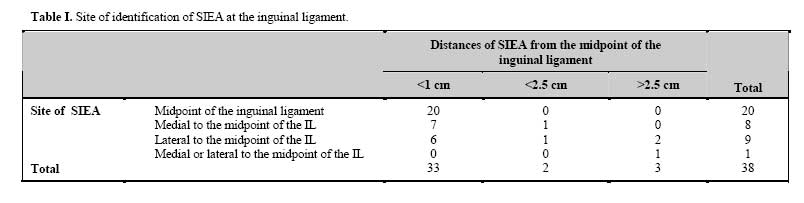
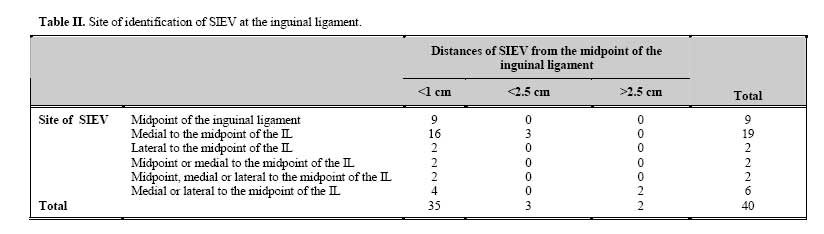
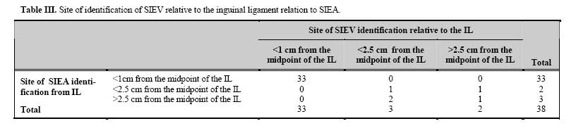
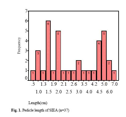
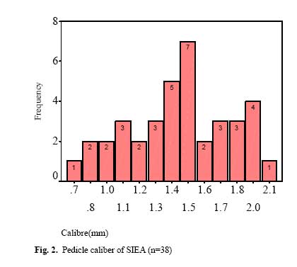
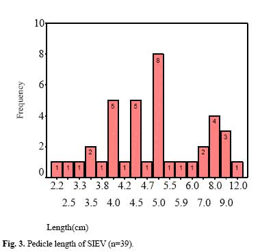
Medical Journal of the Islamic Republic of Iran , Vol. 20, No. 3, November, 2006, pp. 101-106 Anatomy of the Superficial Inferior Epigastric Artery Flap Mahdi Fathi, M.D, Ebrahim Hatamipour, M.D.,* hamid Reza Fathi, M.D., And Ali Pasha Meysamie, M.D., M.Ph. From the Dept. of Plastic and Reconstructive Surgery, Tehran University of Medical Sciences, Imam Khomeini Hospital, Tehran, and the Dept. of Clinical Research, Tehran University, Tehran, Iran. Code Number: mr06023 ABSTRACT Background: Several case studies have described the use of the superficial inferior epigastric artery (SIEA) flap as a pedicled flap for reconstruction of upper and lower ex tremities, or a free fasciocutaneous flap when a large amount of skin coverage is required for hemifacial atrophy, breast or head and neck reconstruction. Apparently, the anatomical findings of previous studies are relatively discrepant. This study was designed to describe the anatomical variations of SIEA and superficial inferior epigastric vein (SIEV). Keywords: Superficial inferior epigastric artery, Free flap, Anatomy INTRODUCTION Since Wood first described the superficial inferior epigastric artery (SIEA) flap as an axial pattern for reconstructing a forearm defect in 1863, several studies have reported its use as a pedicled or free flap for reconstructing upper and lower extremities, head and neck, breast and genital organs.1-6 Early in the 1990s, the free SIEA flap was applied to breast reconstruction. As this flap is supplied solely from the superficial system, it can be elevated from the anterior rectus sheath to transfer abdominal skin and subcutaneous tissue, without harvesting or incisingincising the muscles.7-9 On the other hand, the transverse rectus 7abdominis myocutaneous (TRAM) flap is associated with considerable donor site morbidity, such as abdominal hernia and prolonged hospital stay.10 Moreover, the SIEA flap is particularly advantageous for bilateral autologous tissue breast reconstructions, because the defect at the donor site can be repaired directly, the flap transfer leaves a scar similar to abdominoplasty, and the required superficial dissection at the femoral triangle poses low risk injury to vessels.2, 11 There were no differences in aesthetic results of breast reconstruction using TRAM, deep inferior epigastric artery perforator (DIEP), or SIEA flaps. The disadvantages of the SIEA flap include inconsistent vascular pedicle anatomy and the shorter and smaller diameter vascular pedicle.10 The SIEA is a direct cutaneous vessel that originates from the medial side of the common femoral artery, approximately 2 cm below the inguinal ligament. It pierces the cribriform fascia and passes anterior to the inguinal ligament in a superior course through the subcutaneous tissue of the abdominal wall. The area of skin supplied by the artery is 140±100 cm 2 and tends to run in a curvilinear manner, 5 cm above the iliac crest. The flap extends from the lateral anterior superior iliac spine to the lateral border of the rectus muscle and from the umbilical area to the pubic tubercle.12, 13 The anatomical position of vessels of the SIEA flap have been studied in some clinical case series and limited dissections of cadavers. However, the anatomical findings of previous studies seem relatively discrepant. A recent study has shown that the SIEA is more consistently present and larger in caliber than previously believed.14, 15 This study demonstrated a series of 40 cadaver dissections and described the anatomical variations of SIEA and SIEV by caliber and size of flap pedicle, origin or drainage and position of vessels. Also, the correlation between the size and length of the SIEA pedicle has been described. MATERIAL AND METHODS Anatomical dissection of cadavers A series of 40 dissections were performed on 20 preserved or fresh male cadavers. The cadavers' mean age at death was 39.05±14.06 years. An oblique incision was made overlying the inguinal ligament to dissect the skin and subcutaneous tissue of the lower border of the SIEA flap. An additional small incision (2 cm) was carried out longitudinally below the inguinal ligament to identify the precise site of the vessels. The SIEV or venae comitantes and SIEA positions relative to the midpoint of the inguinal ligament were measured along the course of the vessels. The SIEA passed superiorly and laterally from the femoral triangle to cross the inguinal ligament deep in Scarpa's fascia. Above the inguinal ligament, the artery penetrated Scarpa's fascia to lie in the subcutaneous tissue. The SIEV was found laterally, or medially superficial, or deep in the SIEA. A number of vessels were identified at each of the dissection sites. The sites of vessels identified were medial and lateral to the midpoint of the inguinal ligament. The origin of the SIEA from the femoral artery was identified as a single, common trunk with other arteries or double SIEA. The SIEV or venae comitantes were traced towards the femoral vessels. The drainage of SIEV or venae comitantes into the saphenous bulb or other veins was identified as a single or more than one venous trunk. The external caliber of the SIEA and SIEV was measured using Vernier calipers. In each case, the caliber was measured at the most suitable site for applying microsurgical anastomosis. Also, the caliber of the most suitable vessel trunk was used to calculate the average of the caliber. Measurements of SIEA caliber were considered at the vessel origin from the femoral artery or common trunk with other vessels. SIEV measurements were considered at the drainage to the saphenous bulb or other veins. The caliber of venae comitantes was measured independently. The pedicle length of SIEA was considered as the distance between the most suitable proximal site and its disappearance into the tissue flap at the inguinal ligament. The pedicle length of SIEV was considered as the distance from the drainage site of the vein or venae comitantes to its disappearance into the tissue flap at the inguinal ligament. Data Analysis Descriptive statistics of variables were summarized as a mean ± SD (range) or as percentage of cadavers having 2 the characteristics. For the categorical variables, the statistical significance of difference among the sites of iden-0 tification of vessels was evaluated by using χ2 tests. Linear regression analysis was performed to determine the correlation between size and caliber of the vascular pedicle. The level of P<0.05 was considered as statistically significant. Statistical analysis was performed using SPSS version 11.5 (Chicago, IL, USA). RESULTS Position and origin of vessels From 20 cadavers, the SIEA was identified at the inguinal ligament level in 38 (95%) dissections. The SIEA was absent only in one dissection; in another dissection, it was not identified due to a relatively large hematoma at the groin. Double SIEAs were observed in two distinct cadavers, arising from the common femoral artery (CFA) with a common or double trunk. The origin of the individual SIEA arose directly from CFA, as a common trunk with the superficial circumflex iliac artery (SCIA), as a common trunk with the pudendal artery (PA) and superficial femoral artery (SFA) in 22 (57.9%), 7 (18.4%), 2 (5.3%), and 5 (13.2%) of the 38 dissections, respectively. In the remaining two cases, the SIEA originated from the external iliac artery or the lateral circumflex femoral branch of the deep femoral artery. The SIEA was found within 1 cm of the midpoint of the inguinal ligament in 33 of 38 cadavers. However, in five cases, the SIEA was identified within 1 to 2.5 cm far from the midpoint of the inguinal ligament. In nine cases, the SIEA was found lateral to the midpoint of the inguinal ligament. The remaining 28 SIEAs crossed the inguinal ligament at the midpoint (20 cases) or medially to the midpoint; in one dissection only, two SIEAs crossed the inguinal ligament either medially or laterally to the midpoint (Table I). In all the dissections, the SIEV was identified as an individual vein or pair of venae comitantes. In 24 dissections (60%), the venous drainage was as an individual vein into the saphenous bulb. In the remaining 16 cases, a pair of venae comitantes drained into the saphenous bulb. In some cases, the venae comitantes joined together; in other cases, the SIEV or venae comitantes were joined by other veins. In most dissections, the SIEV or venae comitantes were drained into the saphenous bulb at the midpoint or medially to the midpoint of the inguinal ligament (Table II). In 33 cases, the SIEV and SIEA were found within 1 cm of the midpoint of the inguinal ligament. Thus, the SIEV or venae comitantes were identified close to the SIEA crossing the inguinal ligament (P<0.0001) (Table III). Length and caliber of the vascular pedicle The pedicle length of the SIEA was measured in 37 dissections. The mean±SD length of SIEA was 3.04±1.73 (0.5- 7) cm. The median length of SIEA was 2.5 cm. In 17 (45.9%) of 37 dissections, the pedicle length of SIEA was 3 cm or longer (Figure 1). The caliber of the SIEA was measured in 38 dissections. The mean±SD caliber of SIEA was 1.45±0.35 (0.7- 2.1) mm. The median caliber of SIEA was 1.5 mm (Figure 2). In 20 (52.6%) of 38 cases, the caliber of SIEA was 1.5 mm or longer. In 13 (34.2%) of 38 cases, the caliber of SIEA was less than 1.4 mm. When the SIEA was harvested at the origin from its common trunk with SCIA in seven dissections, the mean caliber and length of pedicle was 1.63±0.28 mm and 4.09±1.79 cm, respectively. When the SIEA originated directly from CFA and SFA, the mean caliber was 1.36±0.41 mm and 1.48±0.08 mm, respectively. The length of the pedicle was 2.5±1.35 cm when the SIEA originated directly from CFA. It was 3.8±2.71 cm when the vessel originated directly from SFA. The pedicle length of SIEV was measured in 39 dissections. It ranged from 2.2 to 12 cm with a mean±SD of 5.45±2.08. The median length of SIEV was 5 cm (Figure 3). The pedicle caliber of SIEV was measured in all dissections. It ranged from 1.6 to 4 mm with a mean±SD of 2.14±.45 mm. The median caliber of SIEV was 2 mm (Figure 4). The linear correlation between the caliber of SIEA (x) and the length of SIEA (y) was expressed by the following equation with r = 0.517 and r 2 = 0.268: Y= 0.517X -0.586. The length of SIEA was increased 2.5 mm for every 0.1 mm increase of the caliber of SIEA. DISCUSSION The SIEA is a direct cutaneous vessel that originates from the medial side of the femoral artery approximately 2 cm below the inguinal ligament. It pierces the cribriform fascia and passes anterior to the inguinal ligament in a superior course through the subcutaneous tissue of the abdominal wall. The flap extends from the anterior superior iliac spine to the lateral border of the muscle, and from the umbilical area to the pubic tubercle.12, 13 However, anatomical findings of previous studies seem relatively discrepant. The anatomical information of the flap was well described by Taylor and Daniel in 1957. In 35 of 100 dissections, the SIEA was absent or could not be identified. The average caliber of the SIEA trunk was 1.4 mm (48%) and 1.1 mm (17%) when the artery originated as a common trunk with SCIA and as an independent vessel, respectively.15 However, the anatomy of consistent veins and length of vascular pedicle was not reported. In Hester et al's study only 1 of 16 SIEA flaps was inadequate for free transfer.16 In the series of 27 free SIEA flaps, Stern and Nahai reported the mean length of the pedicle as 4 cm. In two cases, vein graft was necessary to increase the SIEA length for reconstruction. Arnez et al. described 20 breast reconstructions, using SIEA, DIEP, and TRAM flaps. Based on their protocol, exposure of the SIEA was performed for every patient whether its caliber was equal to or larger than 1.5 mm. The SIEA flap was used successfully in five breast reconstructions. Also, The SIEA was absent in 8 of 20 patients.2, 17 Recently, an anatomical study of 11 cadavers was reported by Reardon et al.13. The SIEA and SIEV were identified in 20 (90.9%) and 21 dissections, respectively. The SIEA originated from CFA within 1 cm of the midpoint of the inguinal ligament in 15 of 20 cases. The average of SIEA length was 5.2 cm, ranging from 3 to 7 cm in 20 dissections. The mean caliber of SIEA was 1.9 mm (1.2- 2.5). The SIEA had a caliber of 1.5 mm or greater in 16 of 20 cases and a pedicle of 5 cm or longer in 14 cases. The venous drainage was as an individual vein, a pair of venae comitantes or both in 12, 8, and 1 dissections, respectively. Except in one case, all the veins drained into the saphenous bulb. The mean caliber and pedicle length of the SIEV was 2.1 mm and 6.4 cm, respectively. Their findings suggested that the SIEA is more constant in its presence and has a greater caliber than previously reported. Also, it may have greater potential for clinical use than previously assumed.13 Chevron conducted a prospective study of 14 SIEA flap breast reconstructions, describing its reliability and limitations. The SIEA flap could not be used in 33 (70%) of 47 reconstructions because its criteria were not met. The SIEA was absent in 24 (51%) cases. In six patients (13%), the SIEA was considered too small for reliable use. The aesthetic results of breast reconstruction by using the SIEA, DIEP, or TRAM flaps were found to be indistinguishable. However, two SIEA flaps that required emergency reoperation were 1.5 mm in diameter.8 Offman et al. studied a series of five cadavers to describe the vascular anatomy of the lateral lumbar region. A dominant SIEA was present in 9 of 10 dissections. Using a technique of arterial injection, the mean length and emerging diameter of SIEA were measured as 96±60 mm and 1.2± 0.4 mm, respectively.12 The authors presented anatomical variations of the SIEA and SIEV in 40 cadaver dissections. In the current study, the SIEA was absent only in one dissection and it could not be identified in another dissection because there was a hematoma at the groin. Also, in all the dissections, the SIEV was identified as an individual vein or pair of venae comitantes. The presence of SIEA vessels was more than what was previously reported. Although two recent series of cadaver dissections had shown that SIEA was present in more than 90% of cases,13, 14 other clinical or cadaveric explorations had dissimilar findings. In our study, the caliber of SIEA was 1.5 mm or greater in more than 50% of the dissections. In clinical studies of the SIEA flap transfer, the pedicle caliber was found to be satisfactory in less than one-third of the patients.2, 8, 11, 15 and 17 However, Reardon et al. reported 80% of the dissections had a caliber of 1.5 mm or greater. Thus, clinical studies have to be undertaken, especially randomized clinical trails of microsurgical transfer of the SIEA rather than other alternatives. Although a diameter of 1.5 mm has been accepted as the lower limit of successful anastomosis in breast reconstruction, these clinical experiences are inadequate. Although the SIEA is 1.5 mm or less in caliber the flap can be appropriate for other microsurgical reconstructions. Recently, the intraoral or extra-orally transferred superficial inferior epigastric artery adipose flap with a short pedicle has been used successfully for three patients with facial contour deformities. Regarding the anatomy of the superficial inferior epigastric artery system, it seems that the ascending branch of the superficial circumflex iliac artery compensates for the superficial inferior epigastric artery deficit. The superficial inferior epigastric artery flap can be raised in all patients, with the superficial inferior epigastric artery itself or with an ascending branch of the superficial circumflex iliac artery and large superficial epigastric vein system. The major disadvantage of a superficial inferior epigastric artery adiposal flap is that fine technical skills are necessary to dissect and anastomose the small and short pedicle vessels for the superficial inferior epigastric artery flap with a short pedicle. However, this flap seems to be the best application for facial contouring surgery and it is indicated especially for children and for young women who expect to become mothers.6 The length of SIEA is correlated to its caliber, that is, we expect to have a longer SIEA with greater caliber during dissections. The SIEA and SIEV are more consistently present than those previously reported. The authors suggest that the SIEA flap can be applied for fasciocutaneous defect repair, potentially in the reconstruction of breast, head and neck, and extensive phalloplasty. ACKNOWLEDGEMENTS The authors wish to thank SPI Publisher Services LLC for assistance with technical support. REFERENCES
Copyright 2006 -Medical Journal of the Islamic Republic of Iran The following images related to this document are available:Photo images[mr06023t2.jpg] [mr06023t3.jpg] [mr06023f3.jpg] [mr06023f2.jpg] [mr06023f1.jpg] [mr06023t1.jpg] |
| |||||||||