 |
search
for |
| About Bioline | All Journals | Testimonials | Membership | News |
|
||||||
|
||||||
Indian Journal of Medical Sciences, Volume 57, Number 8, August 2003, pp. 350-354 Serum biochemical markers in carcinoma breast Late R K Seth, Simmi Kharb, D P Kharb* Department of Biochemistry and *Surgery Pt. B.D. Sharma PGIMS, Rohtak, Haryana.
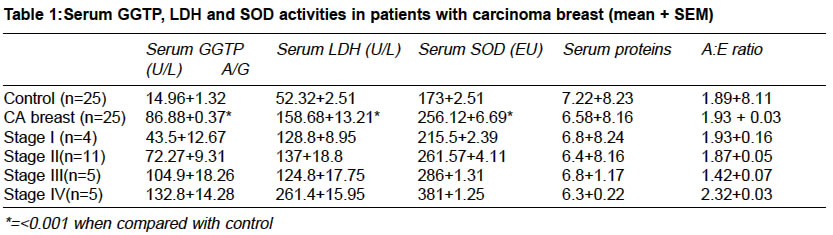
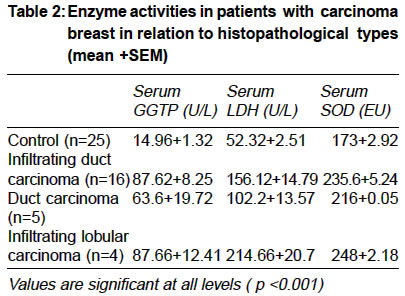
Accepted Date: 10-06-2003 Code Number: ms03013 ABSTRACT Background: Despite the extensive research for many years throughout the world, the etiopathogenesis of cancer still remains obscure. For the early detection of carcinoma of various origins, a number of biochemical markers have been studied to evaluate the malignancy. Aim: To analyse serum gamma glutamyl transpeptidase (GGTP), lactate dehydrogenase (LDH) and superoxide dismutase (SOD) in carcinoma breast patients. Settings & Design: The serum biochemical markers were estimated in twenty five histopathologically confirmed patients with carcinoma breast and equal number of healthy age-matched individuals served as control. Material & Methods: Serum gamma glutamyl transpeptidase (GGTP), lactate dehydrogenase (LDH) and superoxide dismutase (SOD) were estimated and their sensitivity determined. Statistics: Data was analysed with student's `t'-test and sensitivity score of these markers was determined. Results & Conclusions: The mean serum GGTP, LDH and SOD activities in patients with carcinoma breast were tremendously increased as compared to controls, and a steady increase was observed in their activities from stage I through stage IV as well as following distant metastasis. Serum GGTP, LDH and SOD might prove to be most sensitive biomarkers in carcinoma breast in early detection of the disease. Key Words: Gamma glutamyl transferase, Lactate dehydrogenase, Superoxide dismutase, Breast cancer. INTRODUCTION Despite the extensive research for many years throughout the world, the etiopathogenesis of cancer still remains obscure. For the early detection of carcinoma of various origins, a number of biochemical markers have been studied to evaluate the malignancy.1 However, no single marker has proved to be a sensitive and specific indicator of early malignancy. Under the normal conditions, each tissue maintains a steady and consistent enzymatic pattern which is significantly altered in malignancy, because membrane constituents are shed into the surrounding milieu at increased rate when cells replicate more rapidly. The enzymes and proteins present in nucleus, cytoplasm and mitochondria are also released in the circulation when cells are destroyed. The enzymatic changes in malignant tissue may result from genetic reprogramming to malignant behaviour, a likely strategy for survival of tumours. Moreover, the protection of cell against cytotoxic effects of active oxygen species is achieved through superoxide dismutase (SOD), an enzyme shown to protect DNA, proteins and cell membranes from oxidative stress for the survival of the tumour cell, that is expected to be increased.2 Besides, many quantitative alterations in serum protein in patients with cancers of various origins have been elucidated.3 Hence the present study is aimed at providing some of the promising biomarkers, directly associated with breast tumour progression (histopathological types) which ar inexpensive, accurate, identified by easy methods of detection and validated, that may be of some prognostic and diagnostic significance. MATERIAL & METHODS The study was carried out in 25 histopathologically confirmed female patients of carcinoma breast admitted in Surgical Ward of Postgraduate Institute of Medical Sciences, Rohtak (India). Out of twenty five patients, sixteen had infiltrating duct carcinoma, five duct carcinoma, four had infiltrating carcinoma in relation to histopathological findings. Patients with myocardial infarction, jaundice or liver disease, leukaemia, polycythaemia, megaloblastic anaemia, pancreatic disease and diabetes mellitus were excluded. Patients who had already received or were under treatment for malignancy were also excluded. The patients with lump in breast were graded into various clinical staging as per Manchester Classification and equal number of healthy age-matched individuals served as control. Under all aseptic conditions, blood samples were drawn by venepuncture and collection in clean vial,serum gamma glutamyl transpeptidase (GGTP),4 lactate dehydrogenase (LDH)5 and superoxide dismutase (SOD)6 were estimated spectrophotometrically. Total serum proteins, serum albumin and albumin globulin ratio (A: G) were quantitated.7,8 The observations were statistically analysed and student's t-test (unpaired) was applied. The ability to identify true positive cases was calculated by using sensitivity score index. The sensitivity score for GGTP, LDH and SOD was mathematically calculated as below9: Sensitivity score: Number of patients with more
than normal levels of enzyme activity RESULTS The mean serum GGTP, LDH and SOD activities in patients with carcinoma breast were observed to be tremendously increased as compared to controls. Moreover, a steady increase in the activity of GGTP was observed from stage I through stage IV (Table 1). Similar statistically significant increase (p <0.001) in the activity of LDH and SOD were also noticed. The mean serum GGTP activity was lower in duct carcinoma when compared with infiltrating duct or lobular carcinoma (Table 1). It is noteworthy that patients with localised disease (stage I to III) showed about two and a half fold increase in LDH, whereas, five-fold increase was observed in patients with dinstant metastasis (Table 2). The serum SOD ctivity was increased with the progression of the disease (Table 2). The sensitivity score for serum GGTP, LDH and SOD were 75%, 84% and 80%, respectively. The pattern in serum proteins has been shown in Table 1. DISCUSSION Tumour associated markers reflect behavioural changes from tissue to blood, resulting in change in levels of enzymes, enzyme variants, proteins and hormones both in cancerous tissue and blood because of unchecked proliferation of cells.10 Therefore, alteration in particular enzyme contents in serum could be a good index of malignancy in its early and best manageable stage if sufficiently specific and sensitive. Gamma glutamyl transpeptidase (GGTP) is a membrane bound glycoprotein enzyme present in normal human serum. It may be a sensitive indicator of hepatocellular damage and biliary obstruction.11 Howeever, increased serum GGTP activity in patients with carcinoma breast has also been documented.12,13 Much more pronounced increases ( by six-fold) have been noticed in the present study. Moreover, the sensitivity score was 76% for GGTP. Nevertheless, a study increase (from stage I to IV) (Table 1) in serum GGTP activity may indicate it as an excellent biochemical marker. Lower level of this enzyme in sera of patients with duct carcinoma as compared to infiltrating duct or lobular carcinoma (Table 2) could be the consequence of infiltration of growth in the surrounding tissues. Because of infiltrative growth, there may be more leakage of this enzyme into extracellular fluid. To our knowledge such histopathological relationship is lacking in literature. Elevated serum LDH levels in patients with carcinoma breast has been estimated by many workers.14 In the present study, serum LDH levels were significantly higher in carcinoma breast patients than in controls. The patients with localized disease (stage I to III) showed two and a half-fold increase, whereas five-fold increase was observed in patients with distant metastasis. There were two patients with bony metastasis (mean levels 225 and 256 U/L) and two with hepatic metastasis (mean levels 316 and 286 U/L). Thus, serum LDH levels increase enormously once the tumour metastasizes and correlates well with the increase in tumour load as observed by others in the past.15 Infiltrating carcinoma had higher levels of LDH as compared to duct carcinoma. No report is available in literature regarding such histopathological relationship, but, it is generally agreed that serum LDH levels directly correlate with fixity of growth.15,16 Thus, it can be inferred that aggressiveness of the growth directly correlates with serum LDH levels. Also, LDH had a sensitivity score of 84%. So, serum LDH levels may also be of diagnostic as well as prognostic value in patients with carcinoma breast. Quantitative analysis of serum proteins showed hypoproteinemia, hypoalbuminaemia and hyperglobulinaemia. Increase in alpha1 and alpha2 - globulin was observed in electrophoresis. Though the reasons for these changes in neoplastic disease is not well understood, a decrease in albumin concentration is well documented.17 Nonetheless, decrease in levels of albumin as a result of liver damage could only be speculated when secondaries are present in liver. Our results are in agreement with those reported in literature.18 A/G ratio was decreased from stage I through stage IV, stage II showing a 50% decrease as compared to controls. These findings might be important in diagnostic purposes in advanced breast cancers. Therefore, assessment of total and differential levels of serum proteins may be of little significance in early detection of carcinoma breast. There are very few studies available on altered levels of SOD in breast lesion.18 In the present study SOD levels were significantly increased as compared to controls. It was interesting to note that the levels of SOD were significantly higher from stage I to stage IV. In stage IV, the values were markedly increased (1.5 times) than in controls. Thus, a proportionate increase has been observed in SOD activity with progression of malignancy. The release of serum SOD from rapidly multiplying tumour cells may be an effort to protect themselves from oxidative damage. It is therefore concluded that serum GGTP, LDH and SOD might be excellent biomarkers in carcinoma breast and may help in early detection of the disease. Nevertheless, haemoglobin, serum proteins may be altered in the late stage of proliferation. Further work to include a large population inflicted with this disease are underway in our laboratory. REFERENCES
Copyright 2003 - Indian Journal of Medical Sciences. The following images related to this document are available:Photo images[ms03013t1.jpg] [ms03013t2.jpg] |
| |||||||||
