 |
| About Bioline | All Journals | Testimonials | Membership | News |
|
||||||
|
||||||
Indian Journal of Medical Sciences, Volume 57, Number 9, September 2003, pp. 383-386 ALTERATIONS IN THE INGESTIVE BEHAVIOUR OF RATS FOLLOWING UNILATERAL LESIONS IN THE CEA & BLA B GANARAJA, P S JEGANATHAN Department of Physiology
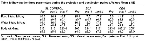
Accepted Date: 30-12-2002. Code Number: ms03020 INTRODUCTION It is well known that the primary control of the ingestive behaviours in the animals rests in the hypothalamic centres. The modulatory activities of the other subcortical areas on this subject had been widely debated. The involvement of the amygdaloid centres in this had been confirmed.1-6 Lesion and stimulation studies involving the amygdala showed that generally the central and dorsal aspects being fecilitatory, while the basal and the lateral portions showed inhibitory influences on the food and water intake in animals.7 In our previous study5 we had demonstrated that the lesions placed in the basolateral nucleus of the amygdala (BLA) increased the food & water intake and the lesions of the Central nucleus (CEA) decreased the intake of both food and water. However it was thought that only the bilateral lesions of the amygdaloid areas would be able to produce these effects. So Gloor8 in his monograph on amygdala suggested that the lesions must be bilateral as far as the amygdaloid areas were concerned. The literature available on the unilateral lesion effects of amygdaloid nuclei is rare. Presently, here we have performed unilateral lesions in the two distinct areas of the amygdala the BLA and CEA. We studied the parameters namely, food intake, water intake and the changes in the body weight of the animals (Wistar albino rats) before the lesion and after the lesion. We observed distinct behavioural pattern in our studies. MATERIAL AND METHODS Wistar strain male albino rats bred in the laboratory (n=21) were used in this study. The animals were housed in standard metallic cages under standard laboratory conditions. The animals were provided with ad lib. access to food (rat feed pellets) and clean water. They were maintained in strict compliance to the animal ethics as per the recommendations of the committee. Stereotaxic surgery was performed to lesion the BLA and CEA unilaterally, in these animals. The rats were anaesthetised by injecting Nembutal (40 mg/kg body wt). The lesions were produced by inserting stainless steel electrode (22 gauge), insulated except for 0.5 mm at the tip. 2 mA DC anodal current was passed for 20 sec. The cathode was connected to the tail. This current was adequate to produce a lesion of about 1mm diameter, which would cover the area of BLA or CEA. First two days after each lesion were left for recuperation. In the sham lesioned control animals only the surgical procedure was performed and no electrical current was passed. The data were collected for two weeks after the recuperation period. The stereotaxic coordinates9: BLA :- AP = - 3 mm posterior to bregma, CEA : - AP = - 2.7 mm posterior to bregma, Premeasured quantities of water, food pellets (BBL India Ltd.) were provided to the rats ad lib. and the quantity consumed per day was measured everyday between 0900 and 1000 hrs. Average consumption per day for 7 days before the lesion (prelesion) and for two weeks after the lesion (postlesion) was measured and tabulated. Mean (±SE) of the daily intake of food and fluids was calculated and represented in the table and figure. Body weight was also measured periodically before and after the lesions (Table 1). At the end of the observation period, these rats were sacrificed and transfused with 10% formol saline transcardially. The brain was dissected carefully and preserved for histological processing. Sections 5m thickness were cut and stained with H&E and examined. Only the data from those rats having at least 70-80% lesions in the respective nuclei were accepted and tabulated for statistical analysis STATISTICS Non parametric statistics applied for the behavioural studies, has been selected for the analysis of the results. Wilcoxon signed rank sum test was applied for the comparison of prelesion Vs postlesion data. Probability p<0.05 was accepted as significant. Mann Whitney `U' test was applied to compare the data from different groups with each other in the respective periods. p<0.01 was considered as statistically significant. RESULTS The food intake, water intake and body weight in the CEA (unilaterally - right side) lesioned rats decreased significantly in the first week following the lesion (p<0.05). However in the second week after the lesion, the food intake as well as the water intake were low when compared to the sham lesioned controls (p<0.01), but compared to their own prelesion intake, the decrease was not statistically significant. The BLA (unilateral) lesioned animals showed significantly increased food intake and body weight in the two weeks following the lesion (p<0.05 for both weeks). Their water intake did not show significant change. DISCUSSION The unilateral lesion of the basolateral nucleus (BLA) showed increased food intake and also the body weight. In our previous report5 we had performed bilateral lesions of BLA and showed that there was increased food intake. This is suggestive of inhibitory role for this centre for ingestive behaviour. Unilateral lesions in the central nucleus (CAE) resulted in the decreased food intake, water intake and the body weight in the two consequent weeks following the lesion. Some reports3,4 suggested the possibility of motor failure to be the cause for the decreased feeding after the lesion of the CEA. This was said to be the result of the damage caused to the fibers traversing the area adjacent to the CEA. But in the previous studies in our laboratory5 we had shown that the discrete lesions in this area do cause the reduced ingestive activites. The reports on the unilateral lesions of the amygdaloid nuclei are rare. Gloor8 had suggested that the lesions in the amygdala had to be bilateral to produce any detectable effects in animals. However, enthused by the findings of our previous study on the bilaterally lesioned animals, we decided to explore the effects of unilateral lesions in the rats. From the present study, we found that unilaterally lesioned animals showed definite alterations in the ingestive behaviours. The BLA lesioned animals showing increased intake and the CEA lesions producing decrease. Although the intensity of these changes were lesser than that we had observed for the bilaterally lesioned animals.5 It is understood that many of the amygdaloid influences on behaviours were exerted via the hypothalamic centres.6 So obviously the effects of lesions in the amygdaloid nuclei will be less striking when compared to the hypothalamic nuclei. Thus the unilateral lesions of amygdala may produce lesser effects than bilateral lesions. We extended our study for only two weeks following lesion. So further study is required to address the other issues regarding this, as to prove whether these effects are temporary or permanent, and also the nature of the activity of the intact side of the brain to mainatain the homeostasis. From the present study, we could conclude that the unilateral lesions of the BLA produce increased food intake and body weight and unilateral lesions of CEA decreased the ingestive activites. This suggested that the inhibitory and excitatory influences of the two amygdaloid centres were evident even after the unilateral lesions contrary to the existing belief that the amygdaloid lesions have to be bilateral to show the effects. REFERENCES
Copyright 2003 - Indian Journal of Medical Sciences. The following images related to this document are available:Photo images[ms03020t1.jpg] |
| |||||||||
