 |
| About Bioline | All Journals | Testimonials | Membership | News |
|
||||||
|
||||||
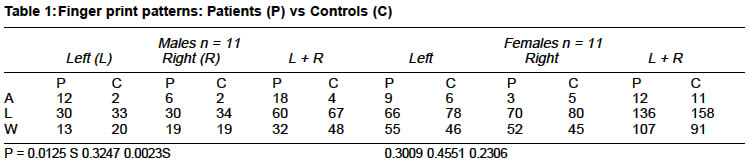
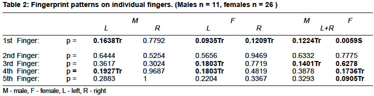
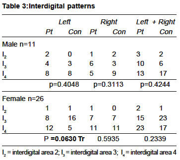
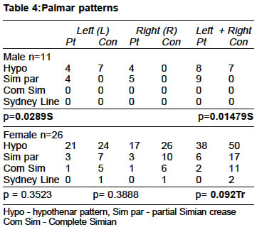
Indian Journal of Medical Sciences, Volume 57, Number 10, October 2003, pp. 437-441 DERMATOGLYPHICS IN RHEUMATOID ARTHRITIS Roopa Ravindranath,# R Shubha,~ H V Nagesh,* Job Johnson,**# Sayee Rajangam***# M.B.B.S., M.S. Associate Professor; *M.B.B.S., M.S., F.R.C.S. Orthopaedician, **B.Sc., Junior Technician; ***M.Sc., Ph.D., Prof. & Head. #Division of Human Genetics, Department of Anatomy St. John's Medical College, Bangalore - 560 034; ~Department of Anatomy, Kempegowda Institute of Medical Sciences, Bangalore; *80 J Block, Broom Field Residence Kennington Road, Willes borough Ashford KENT TN 240LY U.K. Correspondence: Dr. (Mrs.) Roopa Ravindranath, Associate Professor, Department of Anatomy, St. John's Medical College, Bangalore-560034. E-mail: sjmcanat@vsnl.com Accepted Date: 10-06-2003 Code Number: ms03028 ABSTRACT Patients with rheumatoid arthritis have been referred to Division of Human Genetics for counselling. Qualitative dermatoglyphics comprising of finger print pattern, interdigital pattern, hypothenar pattern and palmar crease were studied on 26 female and 11 male rheumatoid arthritis patients. Comparison between patient male and control male; and patient female and control female has been done. `Chi' square test was performed. In male patients, with hands together, arches were increased, loops/whorls were decreased. Partial Simian crease was significantly increased. In the right hand, patterns were increased in the 3rd interdigital area. On the other hand, in female patients there was a significant increase in whorls and decrease in loops on the first finger on both the hands, increase in arches on the 3rd finger; both arches and whorls on the 4th finger of left hand. Present study has emphasized that dermatoglyphics could be applied as a diagnostic tool to patients with rheumatoid arthritis. Key Words: Rheumatoid arthritis, Dermatoglyphics, Palmar crease, Simian crease. INTRODUCTION Rheumatoid Arthritis is a multifactorial condition (polygenic and environmental) and so also the dermatoglyphics (DGs) which is the study of finger print patterns and ridge counts. Factors determining rheumatoid arthritis in-utero may influence dermatoglyphic patterns. DGs could be used as a marker to detect the disease. Hence the following study has been undertaken to detect the presence of correlation between DGs and rheumatoid arthritis. Division of Human Genetics, Department of Anatomy, St. John's Medical College, Bangalore is a referral centre for karyotyping and counselling. Along with chromosomal analysis, the other investigations carried out at Division of Human Genetics are buccal smear and DGs. MATERIAL & METHODS Patients diagnosed in Department of Orthopaedics, St. John's Medical College Hospital, confirmed as having rheumatoid arthritis were referred to Division of Human Genetics for counselling. Patients consisted of 26 females (26-70 yrs) and 11 males (18-54 yrs). Controls were of similar numbers, aged between 45-84 yrs for females and 40-75 yrs for males. Modified Purvis - Smith method1 was applied to obtain the dermatoglyphic prints. Printers ink was smeared on clean dried hands and prints were taken using bond paper and roller. Qualitative dermatoglyphic parameters were studied comprising of finger print patterns, patterns in hypothenar and interdigital (I1, I2, I3, I4) areas. Hands were studied together and separately. Analysis was done on individual fingers also. Statistical analysis was done using `Chi' square test. RESULTS The observations have been tabulated. Table 1: Finger print patterns - Males: When all the fingers were taken into consideration with hands together, as well as in the left hand, arches were more and loops were less and whorls were significantly less in male patients compared to controls. Finger print patterns - Females: No significant finding was found. Table 2: Finger print patterns on individual fingers - Males: With hands together, the first and third fingers showed a trend towards significance with an increase in arch and decrease in whorls in both the fingers and decrease in loops in the 3rd finger. Further analysis with hands separate showed that there was a trend towards significance in the left hand on the first finger. Similar finding was found on the left hand in the 4th finger of males. Finger print patterns on individual fingers - Females: With hands combined, significant finding was present with increase in whorls and decrease in loops on the 1st and 4th fingers; absence of double loop whorl (DLW) on the 1st finger and arch on the 5th finger. Further analysis, with hands separate showed that a trend towards significance was found in the left hand, for a decrease in loops and increase in whorls on the 1st and 4th fingers and decrease in loop on 3rd finger. Increase in arch was found on 3rd and 4th fingers and DLW was absent on the first finger of the left hand. On the first finger of right hand decrease in loops and increase in whorls was found in female patients. Table 3: Interdigital patterns - Males: Patterns were more in interdigital area I3 and less in I4 with hands combined and in the right hand of male patients compared to controls but it was not of statistical significance. Interdigital patterns - Females: With hands combined there was an increase in patterns in I4 and decrease in I3 in female patients, when compared to controls (P=0.2339) but it was not of statistical significance. Further analysis, however, showed similar trend towards significance in the left hand of female patients (P=0.0630). Table 4: Palmar patterns - Males: Patterns were more in right hand in hypothenar area. Partial Simian crease was significantly increased in both hands separately and with hands combined when compared to controls. Complete Simian crease and Sydney line were absent. Females - Simian crease both partial and complete showed a trend towards decrease with hands combined. Hypothenar pattern was less and Sydney line was absent (p=0.0928) when compared to controls. DISCUSSION Few studies are available on DGs and rheumatoid arthritis. Finger print patterns: (Tables 1 and 2) Taneja et al2 have reported an increase in arches on both the hands and decrease in ulnar loops on the right hand of the female patients with rheumatoid arthritis. In the present study, there was an increase in whorls and decrease in ulnar loops on both the hands and increase in arches on the left hand of the female patients. However, it was not of statistical significance. In male patients, Taneja et al (1993) found increased frequency of whorls on the right hand, increased frequency of arches on the left hand and decreased frequency of loops on both the hands. In the present study, in the male patients, arches were more, loops were less and whorls were significantly less on both the hands together and on the left hand compared to controls. In the study done by Taneja et al (1993) the differences found between male versus female patients were increase in whorls on both the hands and arches on the left hand of male patients; increase in ulnar loops on both the hands and arches on the right hand of the female patients. In the present study, for the male patients, arches were increased on both the hands and ulnar loops were increased only on the left hand. For the female patients, radial loops and whorls were increased on both the hands and ulnar loop on the right hand. Further analysis of individual finger patterns in females showed significant increase in whorls on 1st and 4th fingers with hands together. With hands separate increase in whorls was found. In male patients, with hands together, the 1st and 3rd finger showed a trend towards significance with an increase in arch and decrease in whorls; and decrease in loops on the 3rd finger. In male patients with hands separate there was a similar finding with increase in arch and decrease in loops on 1st, and 4th fingers of left hand. Published reports about the frequency of patterns on individual fingers were not available.
Palmar Patterns In the present study, the left hand of female patients showed a trend towards increase in I4 pattern. No significant pattern emerged for the right hand. Taneja et al (1993) found increase in I3 pattern on both the hands of female patients but it was not of statistical significance. In the present study, comparing male versus female patients, for the right hand, patterns were significantly more in I3 and left hand was not significant in male patients. For the female patients, all I2, I3 and I4 were less. Taneja et al (1993) found increased pattern in I2and I3 for both the hands of male patients whereas female patients showed increased I4 pattern in both the hands compared to male patients. Hypothenar pattern: (Table 4) Study of hypothenar pattern showed that 14.28% out of a total of 52 palms had ulnar loop pattern in female patients whereas none of the controls had pattern on hypothenar area. Taneja et al2 found an increased frequency of hypothenar patterns in female patients on both the hands compared to controls. In the present study, patterns were less in hypothenar area in both male and female patients compared to controls. In female patients, Taneja et al (1993) found increased pattern in both the hands of the female patients and in the left hand of male patients compared to controls. In the present study, in female patients, both partial and complete Simian crease showed a decrease with hands together, whereas in males patients, partial Simian crease was significantly increased in both hands separately and with hands together when compared to controls. Complete Simian crease was absent in male patients and Sydney line was absent in both male and female patients. In the present study, comparing male versus female patients, partial Simian crease was increased in male patients and complete Simian crease was increased in female patients in both the hands. Sydney line and Simian crease have not been reported in literature, hence could not be discussed. SUMMARY To conclude, arch pattern on the 1st and 4th finger of the left hand in male patients and whorl on 1st finger of either hand in female patients, could be used as dermatoglyphic markers of rheumatoid arthritis. REFERENCES
Copyright 2003 - Indian Journal of Medical Sciences. The following images related to this document are available:Photo images[ms03028t3.jpg] [ms03028t2.jpg] [ms03028t1.jpg] [ms03028t4.jpg] |
| |||||||||
