 |
| About Bioline | All Journals | Testimonials | Membership | News |
|
||||||
|
||||||
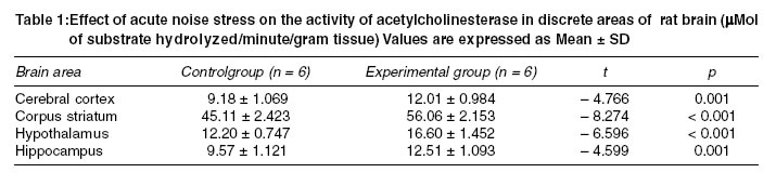
Indian Journal of Medical Sciences, Volume 57, Number 11, November 2003, pp. 487-492 Effect of acute noise stress on acetylcholinesterase activity in discrete areas of rat brain K Sembulingam, Prema Sembulingam, A Namasivayam* School of Health Sciences, University Science Malaysia, Kubang Kerian, Kelantan, 16150, Malaysia; and *Department of Physiology, Dr. ALM. PG Institute of Basic Medical Sciences, University of Madras, Taramani, Chennai-600113. India. Correspondence: Dr. K. Sembulingam, School of Health Sciences, University Science Malaysia, Kubang Kerian, Kelantan, 16150, Malaysia. E-mail: ksembu@yahoo.com Accepted Date: 05-11-2003 Code Number: ms03036 ABSTRACT Effect of various stressor agents on the adrenergic system in brain had been studied extensively. However, reports on the effect of stress on various parameters of central cholinergic system are scanty. And very little is known about the effect of noise stress on the cholinergic system in brain. Hence, it was decided to elucidate the effect of acute noise stress on the activity of the enzyme acetylcholinesterase in discrete areas of brain in albino rats. Male albino rats of Wistar strain were subjected to acute noise stress for 30 minutes. The noise of pure sine wave tone was produced by using a function generator and was amplified. The frequency of noise generated was 1 kHz and the intensity was set at 100 dB. The total acetylcholinesterase activity was determined in the tissues of cerebral cortex, corpus striatum, hypothalamus and hippocampus of brain in these rats. The enzyme activity was estimated by colorimetric method using acetylthiocholine iodide as the substrate. The values were compared with the enzyme activity in the control rats. The activity of the enzyme increased significantly in all the four regions of the brain in rats after exposure to noise stress for 30 minutes. The results of the study indicate that the exposure to acute noise stress could modulate the cholinergic system in these areas of brain in rat. KEY WORDS: Acetylcholinesterase, Brain, Cholinergic system, Noise, Stress, Rat. INTRODUCTION Pioneer stress physiologists like Selye1 had documented most of the stress induced physiological changes. Many processes intervene between stressful stimulus and subsequent responses in the body. A variety of neurochemical reactions are also involved during these processes both in centraland autonomic nervous system. Acetylcholinesterase (AchE) is an important cholinergic enzyme that plays a key role during these processes.2 Extensive studies have been reported on the changes in adrenergic system in brain after exposure to stress.3,4 But, little is known about stress-induced changes in the central cholinergic system. Recent investigations indicate that stress conditions could modulate the central cholinergic system in animals.5,6 And few reports are available to show the changes in the activity of AchE in discrete areas of brain in animals induced by various stress agents like electric shock,7-9 cold stress5 and immobilization or restraint stress.6,10 However, some controversy exists in the results of these studies. The activity of AchE in various areas of brain in animals was noticed to increase,5,7,10 decrease4,8 or remain unaltered11 after exposure to stress. Also, the reports regarding the effects of noise stress on neurological system are very scanty. Noise in public places especially in occupational environments is considered as health hazard. The problem is more pertinent, particularly in occupational settings where exposure to noise for short periods is unavoidable. The relationship between noise and central cholinergic system in animals has not been studied much. Lai (1987) reported the changes in high affinity choline uptake in brain regions induced by 30 minutes exposure to noise (100 dB).12 The effect of noise on the receptors mediating the central cholinergic activity was noticed in discrete areas of rat brain.13 Recently we have observed the reduction in the acetylcholine content in discrete regions of rat brain after exposure to acute noise (1 kHz; 100 dB) stress.14,15 It was thought that the determination of effect of noise stress on AchE activity would be essential to understand the mechanism of noise induced changes in the acetylcholine content in discrete areas of brain. The perusal of literature reveals that, so far, no attempt was made to study the effect of exposure to noise stress on the activity of the enzymes in cholinergic system in whole brain or in discrete areas of the brain in animals. Hence, in this study an attempt was made to elucidate the effect of acute noise stress on the activity of AchE in discrete areas viz. cerebral cortex, corpus striatum, hypothalamus and hippocampus of brain in albino rats. MATERIAL AND METHODS Experimental protocol Stress procedure Extraction and estimation of AchE activity The tissues of each brain region were homogenized separately in a Potter Elvehjem homogenizer by using 0.1 M phosphate buffer (pH=8) at a temperature of 00C. The homogenate was centrifuged at 10,000 g for 5 minutes at 40C. The total acetylcholinesterase activity in the aliquot of the homogenate was estimated by the method followed by Ellman et al.18 The aliquot was mixed with phosphate buffer (pH=8). To this, the substrate acetylthiocholine iodide (Sigma, USA) and dithiobisnitrobenzoic acid (DTNB) reagent (Sigma, USA) were added. Acetylthiocholine iodide was hydrolyzed to thiocholine and acetate by AchE. Thiocholine reacted with DTNB reagent to produce a yellow color. The rate of color development is the measure of the AchE activity. A kinetic profile of the enzyme activity was studied spectrophotometrically at 412 μm at the interval of 15 seconds. The enzyme activity is expressed as the μMol of substrate hydrolyzed/minute/gram tissue. Assay of enzyme activity in each sample was run in duplicate. Statistical analysis of the results was carried out by the software SPSS 11.0 using Student's `t' test to compare the differences between control and experimental groups. RESULTS The data of results are given in the Table 1. Data are presented as means ± S.D of six animals per group. The base line values for the activity of AchE in discrete areas of rat brain in this study were in agreement with the values reported by other investigators.8 The exposure to noise stress for 30 minutes resulted in significant elevation of the enzyme activity in all the four areas of the brain viz. cerebral cortex, corpus striatum, hypothalamus and hippocampus. DISCUSSION Noise is generally considered as a common and strong environmental stress factor for both human beings and animals. Several reflex responses in the body, which are evoked by the exposure to noise stress become more pronounced if the noise is of unknown character or unexpected. The non-familiar noise can produce a fully developed defense alarm reaction with hemodynamic, hormonal, behavioral and various other changes leading to fight and flight reactions.1 Estimation of the activity of AchE was thought worth doing in this study because this enzyme is considered as the classical enzyme in the cholinergic neurohormonal system. It plays an important role in cholinergic neurohormonal system particularly during stress conditions.2 It is established that AchE inhibits the prolonged action of the cholinergic neurotransmitter acetylcholine by hydrolyzing it.19 Also, AchE ranks as one of the most efficient enzymes, since one molecule of the enzyme can hydrolyze 5000 molecules of acetylcholine per second.20 AchE was found to be present in all the areas of cholinergic system in brain, which contain acetylcholine. Of course it is reported to be present also in the brain areas, which are devoid of acetylcholine.21 Among the brain regions, cerebral cortex, corpus striatum, hypothalamus and hippocampus were chosen for investigation in this study because of few interesting facts. First, it was established that only these four regions of brain contained highest amount of cholinergic innervations.8 Second, it was known that the activity of AchE was very much related to the content of total acetylcholine in these four areas of the brain.8,11 Also, the cholinergic neurons in these areas of brain play some specific and unique role in modulating various physiological, biochemical and behavioral responses particularly during exposure to stress.22 The cholinergic nerve fibers in cerebral cortex are involved in cortical activation and selective awareness that are necessary for information and acquisition. The corpus striatum was reported to be extremely rich in acetylcholine content and AchE activity than any other part of brain in animals, particularly rats.8 Functionally, the cholinergic fibers of corpus striatum are responsible for the extrapyramidal control of all the activities.8 The involvement of cholinergic fibers in hypothalamus in stress conditions had been well established. Hypothalamus is known for its role in the commencement and maintenance of various reactions during general adaptation syndrome by activating both sympathetic adrenomedullary and pituitary adrenocortical pathways. Hippocampus is involved in mediating various mechanisms concerned with adaptation, habituation in novel environment and behavioral responses, especially during stress conditions.22 The result of the present study i.e., the elevation of AchE activity in all the four areas of rat brain viz. cerebral cortex, corpus striatum, hypothalamus and hippocampus after exposure to 30 minutes noise (10,000 Hz; 100 dB) stress supports the results of our previous studies. Earlier we had noticed the significant reduction in the acetylcholine content of the same four areas of brain in rats exposed to 30 minutes noise (10,000 Hz; 100 dB) stress.14,15 In view of the results of our previous studies and the present study, it could be speculated that the fall in acetylcholine content in discrete areas of brain in animals exposed to acute noise stress might be due to the release of acetylcholine from the cholinergic nerve terminals and its subsequent hydrolysis by AchE. Literature reveals this type of inverse relationship between the activity of AchE and the content of acetylcholine in brain regions of animals subjected to few other types of stress agents like cold stress5 and electric shock.9 It was not known whether the changes in the activity of the AchE and acetylcholine content in different regions of brain after exposure to acute noise stress could be beneficial or harmful to the organism. But, the exposure to noise could lead to more detrimental behavioral and physiological functions by modulating central cholinergic system in the brain of the organism. The neural mechanism, through which the noise stress could affect the activity of cholinergic system in brain is also not known. Because of the nonexistence of direct connections of central auditory pathway with hippocampus, hypothalamus, striatum and different lobes of cerebral cortex other than temporal lobe, the effect of noise could be an indirect one. Perhaps, the arousal effects of noise could influence the reticular formation, which in turn might modulate the cholinergic pathway.12 So, further investigations are essential to understand the mechanisms involved in the noise induced changes in the central cholinergic system. REFERENCES 1. Selye H. History and present status of stress concept. In: Handbook of stress. In: Goldberger L, Breznitz, editors. London: Macmillan Publishers; 1986. pp 7-8. 2. Appleyard ME. Secreted acetylcholinesterase; non-classical aspects of a classical enzyme. Trends in Neuroscience 1992;15:485-90. 3. Stone EA. Stress and catecholamines. In: Friendhoff AJ, editor. Catecholamines and behavior. New York: Plenum Press; 1975. Vol. 2 pp. 31-72. 4. Kobayashi RM, Palkovits M, Kizer JS, Jacobowits DM, Kopin IJ. Selective alterations of catecholamines and tyrosine hydroxylase activity in the hypothalamus following acute and chronic stress. In: Usdin E, Kvetnansky R, Kopin IJ, editors. Catecholamines and stress. Oxford: Paragon Press; 1976. pp 29-38. 5. Fatranska M, Budai D, Oprsalova Z, Kvetnansky R. Acetylcholine and its enzymes in some brain areas under stress. Brain Research 1987;424:109-14. 6. Fatranska M, Kiss A, Oprsalova Z, Kvetnansky R. Acetylcholinesterase and choline acetyltransferase activity in some hypothalamic nuclei under immobilization stress in rats. Endocrinol Exp 1989;23:3-10. 7. Appleyard ME, Green AR, Smith AD. Acetylcholinesterase activity in regions of the rat brain following convulsion. J Neurochem 1986;46:1789-93. 8. Geoffroy M, Tvede K, Christensen AV, Schou JS. The effect of imipramine and lithium on `Learned helplessness' and acetylcholinesterase in rat brain. Pharmac Biochem Behav 1991;38:93-7. 9. Singh HC, Singh RH, Udupa KN. Electric shock induced changes in free, bound and total acetylcholine and acetylcholinesterase in different brain regions of rats. Indian J Expt Biol 1979;17:304-6. 10. Romero-Vecchione E, Fatranska M, Kvetnansky R. Acetylcholinesterase activity in several hypothalamic and brain stem nuclei after acute and chronic immobilization stress in rats. Endocrinol Exp 1987;2:159-65. 11. Hata T, Kita T, Higashiguchi T, Ichida S. Total acetylcholine content and activities of cholineacetyltransferace and acetylcholinesterase in brain and duodenum of SARTstressed (Repeated cold stressed) rat. Japan. J Pharmacol 1986;41:475-85. 12. Lai H. Acute exposure to noise affects sodium - dependent high - affinity choline uptake in the central nervous system of the rat. Pharmacol Biochem Behav 1987;28:147-51. 13. Lai H, Carino MA. Opioid receptor subtypes mediating the noise-induced decreases in high-affinity choline uptake in the rat brain. Pharmacol Biochem Behav 1992;42:553-8. 14. Sembulingam K, Sembulingam P, Namasivayam A. Effect of acute noise stress on cholinergic neurotransmitter in corpus striatum of albino rats. Pharmaceutical Sciences (Lond) 1996;2:241-2. 15. Sembulingam K. Prema Sembulingam and Namasivayam A. Effect of acute noise stress on acetylcholine content in discrete areas of rat brain. J Environ Biol 1999;20:289-92. 16. Dickens W. In: William IG, editor. Methods of Animal experimentation. New York: Academic press;1973. Vol. IV. pp 47-98. 17. Glowinski J, Iverson LL. Regional studies of catecholamines in the rat brain. I. The disposition of [3H]norepinephrine, [3H]dopamine and [3H]dopa in various regions of the brain. J Neurochem 1966;13:655-9. 18. Ellman GL, Courtney KD, Andres Jr. V, Feather Stone RM. A new and rapid colorimetric determination of acetylcholinesterase activity. Biochem Pharmacol 1961;7:88-95. 19. Collier B. Biochemistry and physiology of cholinergic transmission. In: Brookhart JM, Mount Castle VB, Kandel ER, Gieger SR, editors. Handbook of Physiology. U.S.A: American Physiological Society; 1977. pp. 463-602. 20. Cooper JR, Bloom FE, Roth RH. In: Acetylcholine in the biochemical basis of neuropharmacology. London: Oxford University Press; 1986. pp. 173-202. 21. Butcher LL, Wolf NJ. Histochemical distribution of acetylcholinesterase in central nervous system. Clues to the localization of cholinergic neurons. In: Bjorklund A, Hokfeet Kuhar MJ, editors. Handbook of chemical neuroanatomy. Amsterdam: Elseviar; 1984. Vol. 3. pp. 1-5. 22. Pepeu G. Brain acetylcholine: An inventory of our knowledge on the 50th anniversary of its discovery. Trends in Pharmac Sciences 1983; 4:416-8. Copyright 2003 - Indian Journal of Medical Sciences. The following images related to this document are available:Photo images[ms03036t1.jpg] |
| |||||||||
