 |
| About Bioline | All Journals | Testimonials | Membership | News |
|
||||||
|
||||||
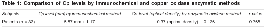
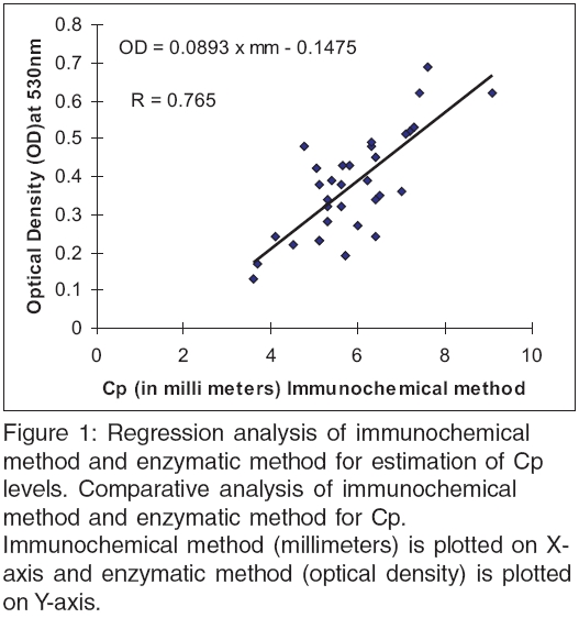
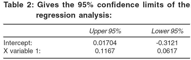
Indian Journal of Medical Sciences, Vol. 60, No. 9, September, 2006, pp. 371-375 ORIGINAL CONTRIBUTIONS Pros and cons of immunochemical and enzymatic method in the diagnosis of Wilson's disease Gnanou JV, Thykadavil VG, Thuppil V University of Seychelles American Institute of Medicine, MAHE, Seychelles Correspondence Address:C/o Dr. Vinod George, Dept. of Biochemistry, St. John's Medical College, Koranamgala, Bangalore - 560 034, India Email: gnanou_j@yahoo.com Code Number: ms06053 Abstract BACKGROUND: Immunochemical method of measuring Ceruloplasmin (Cp) levels for the diagnosis of Wilson's disease has replaced enzymatic method for the main reason of being more sensitive and quantitative.SETTINGS AND DESIGN: In this study, we compared both the methods for various factors such as sensitivity, specificity and the time consumed in the diagnosis of Wilson's disease. MATERIALS AND METHODS: Serum samples from patients (n=33) with a provisional diagnosis of Wilson's disease were analyzed for Cp levels by enzymatic copper oxidase method and immunochemical method using polyclonal antibodies specific to Cp embedded in agar. STATISTICAL ANALYSIS: Pearson's regression analysis was performed to compare the two methods. RESULTS: The mean Cp obtained by immunochemical method is 5.87 mm ± 1.17 and by enzymatic method, it is 0.37 (Optical Density) ± 0.136. Pearson's Regression analysis of the measurements showed a good correlation with an 'r' value of 0.765 between the two methods. CONCLUSION: A good correlation indicated that these two tests are comparable and thus both these methods can be used together for a definitive and better diagnosis of Wilson's disease. Keywords: Ceruloplasmin, copper oxidase method, immunochemical method, Wilson′s disease Cp is a transport protein for copper and is secreted as apoCp. This protein takes up six to eight atoms of copper through a copper transport mechanism using ATP, producing holoCp.[1] The disruption of the copper-transporting mechanism due to mutations (ATP7B) leads to a reduced excretion of copper and accumulation of copper in hepatolenticular cells causing Wilson′s disease. More than 200 known mutations have been characterized and documented.[2] High level of suspicion in pediatric populations presenting with neuro-hepatic problems prompts clinicians to monitor their levels of Cp. The diagnosis of Wilson′s disease is made by measuring the Cp levels, which will be lower, along with clinical signs and elevated liver enzymes.[3] There are two methods for estimating Cp in blood: copper oxidase method and immunochemical method. Copper oxidase method is an enzymatic method and estimates the holoCp, while the immunochemical method estimates the combined apoCp and holoCp. Hence it is more appropriate to estimate the holoCp levels rather than apoCp and holoCp together in the diagnosis of Wilson′s disease. Also, since Cp is an acute phase reactant protein, inflammation due to any cause can increase the Cp levels. Thus immunochemical method has lower specificity than copper oxidase method for the diagnosis of Wilson′s disease.[4] Hence using immunochemical method in patients with Wilson′s disease presenting with inflammation or infection, normal or high Cp levels can be obtained.[5],[6] This problem is overcome by using copper oxidase method, which measures only the holoCp levels. However, copper oxidase method is a qualitative method and has low sensitivity.[7] The aim of this study was to study the pros and cons of these two methods, immunochemical method and copper oxidase method, in patients suspected with Wilson′s disease. Materials and Methods Subjects This study was a prospective study and was conducted over a period of 6 months-from January to June 2005. The study population was selected using an inclusion criterion of pediatric patients between 1 year and 15 years of age, with nonspecific liver diseases in whom Wilson′s disease was the differential diagnosis. Patients with specific liver diseases such as acute or chronic hepatitis and adult patients were excluded from the study. Since this study was restricted to a period of 6 months, all patients who fitted the inclusion criteria were selected for the study. Thus the size of the study population was 33. Of these 33 patients, 31 were male and 2 were female. Two milliliters of blood was collected by standard venipuncture procedure and samples were immediately centrifuged at 5,000 rpm at 25°C and serum was separated. The samples were collected on different days for a period of 6 months as and when patients were recruited. The serum samples were aliquoted into three vials. The first two vials were coded and they were immediately analyzed for Cp levels using both the immunochemical method and the copper oxidase method. The third serum vial was immediately stored at - 20°C for future and repeat analysis. The lab staff members were blinded for this analysis. Ethical clearance Ethical approval was obtained from the Institutional Ethical Review Board of St. John′s Medical College, Bangalore and all subjects gave informed consent. Sample analysis Immunochemical method was done using polyclonal antibodies specific to Cp embedded in agar (Biocientifica, SA Buenos Aires, Argentina). In this method, 10 µL of serum sample was added into the sample well in the agar. The sample was incubated in the agar medium at room temperature for about 12 h. After the incubation period, the Cp concentrations were calculated by measuring the diameter of the radial immunodiffusion by a calibrated scale.[8] This measurement was then compared to a control run along with the sample and result given in mg/dl. The enzymatic oxidase activity of Cp was assayed using para-phenylenediamine in sodium acetate as substrate and the purple product was measured spectrophotometrically at 530 nm.[6],[8] In this method, 100 μL of serum sample was taken in a test tube and incubated with para-phenylenediamine in sodium acetate buffer for 15 min. After the 15-min incubation period at room temperature, the color produced was measured as optical density at 530 nm using a spectrophotometer. This value was then compared to a control/normal sample run along with the test with same lab conditions. If the optical density of the test was lower than the normal, then the test was interpreted as Cp deficiency. If the optical density was same or higher than the control/normal sample, the test was interpreted as normal Cp levels. The within-run CV% and between-run CV% of immunochemical method was 1.57 and 2.63% respectively and the within-run CV% and between-run CV% for copper oxidase enzymatic method was 3.23 and 7.01% respectively. The lower limit of detection of immunochemical is 7 mg/dl. Statistical analysis Analysis was performed with SPSS (SPSS 10.1 for windows, SPSS Inc 2000). The two methods were compared using Pearson′s regression analysis. For regression analysis, 'r' value greater than 0.5 was considered as good correlation. Results The mean Cp obtained by immunochemical method was 5.87 mm ± 1.17 and by enzymatic method, it was 0.37 (optical density) ± 0.136. Pearson′s regression analysis of the measurements showed a good correlation with an 'r' value of 0.765 between the two methods [Table - 1]. From the total observations, the predicted and residual values were plotted onto the X and Y axis to obtain a line-fit plot. Using this plot, a predictive equation was arrived at. For this study, the predictive equation is as follows: Optical density by enzymatic method = 0.0893 x millimeters measured by immunochemical method - 0.1475 [Figure - 1]. The 95% confidence limits of the regression analysis is given in [Table - 2]. Discussion In this study, we found a good correlation between the two methods - immunochemical method and enzymatic method - for the diagnosis of Wilson′s disease. The gold standard method for the diagnosis of Wilson′s disease is through mutation analysis. The only drawback of this method is the cost-effectiveness.[2] Mutation analysis costs approximately US $ 200-250, while the immunochemical method costs approximately US $ 10 and enzymatic method costs approximately US $ 2. Immunochemical method is a sensitive and quantitative method but measures the total Cp (holo + apo Cp). Thus in patients with Wilson′s disease presenting with infection,[9] this test will give a false negative result. On the other hand, copper oxidase method is specific for the holoCp and does not measure the nonbound Cp. But this test has a disadvantage of being less sensitive and being a qualitative test. This test also is labor intensive and time consuming. Thus most labs prefer to use a faster, quantitative and easier method, which is the immunochemical method, for the estimation of Cp. We also found in our hospital through personal communications that clinicians were more inclined to trust a quantitative result than a qualitative one. Hence when using Cp assays for diagnosing Wilson′s disease, the clinicians should be aware of the nonspecificity of the immunochemical method. Moreover, the lab should also take responsibility of informing the clinician about the method used and the advantages and disadvantages of the method. With due consideration of the above points, we analyzed Cp levels in patients with differential diagnosis for Wilson′s disease by these two methods to see if they were comparable. We found that these two methods are comparable. Since they are comparable, both the methods can be used simultaneously. Thus by combining both these methods, the problem of sensitivity and specificity can be avoided. The main drawback of this study was that the patients were not categorized based on inflammatory status. Also a definitive diagnosis of Wilson′s disease using mutation analysis would have given better information of the usefulness of these assays in the diagnosis of Wilson′s disease. The other main drawback of this study was that this study was restricted to pediatric population. In future, perhaps, it may be possible to include all the age groups in order to eliminate the role of age and sex in these assays. To conclude, we found that enzymatic assay is the test of choice for the diagnosis of Wilson′s disease, but combination of both immunochemical and enzymatic method would eliminate the disadvantages of both the methods. Acknowledgment All authors appreciate the efforts put in by Ms. Geetha S. for providing technical help in this study.References
Copyright 2006 - Indian Journal of Medical Sciences The following images related to this document are available:Photo images[ms06053f1.jpg] [ms06053t1.jpg] [ms06053t2.jpg] |
| |||||||||
