 |
| About Bioline | All Journals | Testimonials | Membership | News |
|
||||||
|
||||||
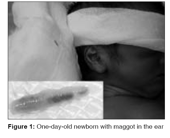
Indian Journal of Medical Sciences, Vol. 62, No. 4, April, 2008, pp. 164-166 Letter To Editor Aural myiasis in a 1-day-old neonate Jain Shraddha, Audhya Arijit, Madhupriya, Nagpure PS Department of Otorhinolaryngology and HNS, Mahatma Gandhi Institute of Medical Sciences, Sewagram, Wardha, Maharashtra Code Number: ms08030 Sir, A full-term neonate girl was delivered by vaginal route in the septic labor room of our hospital. Her birth weight was 1500 g. She was kept in the neonatal intensive care unit. On next day of birth, she developed blood-tinged discharge from her right ear, with incessant cry. On cleaning with a cotton bud, a live 6-mm-long cylindrical white maggot was evacuated along with debris. On turpentine oil instillation, two more white maggots 5- to 6-mm long crawled out of the ear cavity [Figure - 1]. Turpentine oil-soaked cotton pledget was placed at the opening of external auditory canal. No further maggots came out. The maggots were sent to the department of zoology and grown into flies of family Calliphoridae and genus Calliphora, and the species identification was done by the presence of the posterior spiracles on the last abdominal segment of third-stage larva. Facility for species identification by DNA methods was not available with them. Treatment comprised of parenteral ampicillin and cefotaxime; and instillation of Ofloxacin ear drops after removal of maggots. Right ear examination on the seventh day revealed a small healing perforation in the anteroinferior quadrant of the tympanic membrane. The baby was doing well. The mother had presented with septicemia with jaundice. Her WBC count was 26,000/cmm; serum bilirubin, 16 mg/dL; AST, 854 IU/L; ALT, 759 IU/L; ALP, 369 IU/L. Her cervical swab showed pus cells, and culture sensitivity showed heavy growth of Klebsiella sp sensitive to amikacin. Findings of examinations of other systems and laboratory investigations were normal. Myiasis in the neonatal period is rare and almost exclusively found in tropical areas. It is more common in children less than 5 years of age and with a rural background. [1] Flies (Diptera) of various kinds are mostly responsible for myiasis. The fly that caused infestation in our case was a species of blowfly (order Diptera, suborder Cyclorrhapha, and family Calliphoridae). Flies in the family Calliphoridae are generally referred to as ′blowflies′ or ′bluebottles.′ Maggots are classified into three ′instar′ stages. An instar I is about 2- to 5-mm long and corresponds to an age of 2 to 3 days (for average houseflies or bluebottles) from the time the eggs are laid. Myiasis can be classified depending on the condition of the involved tissue, into accidental myiasis, semispecific myiasis, and obligatory myiasis. [2] The majority of cases of human myiasis involve fly species that are facultative parasites; humans represent only a target of opportunity, presented through neglected wounds or lack of sanitary measures. [3] When larvae affect undamaged skin, it is obligatory myiasis. In the present case, this appears to be an obligatory parasitism as the neonate was delivered in an institution and kept in a neonatal ICU after birth. Maggots were removed on the next day of birth, so the larva was instar I. There are very few reports of neonatal myiasis. [4],[5] This case is interesting to report as it is probably the first case of neonatal myiasis in a 1-day-old baby delivered in an institution. References
Copyright 2008 - Indian Journal of Medical Sciences The following images related to this document are available:Photo images[ms08030f1.jpg] |
| |||||||||
