 |
| About Bioline | All Journals | Testimonials | Membership | News |
|
||||||
|
||||||
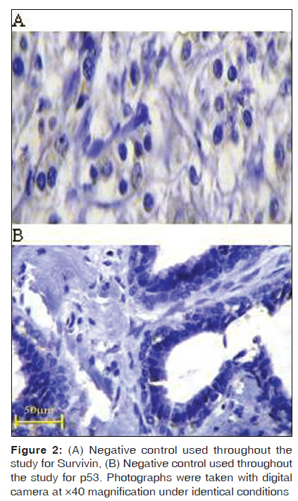
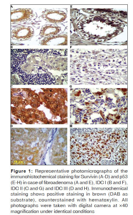
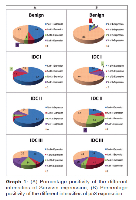
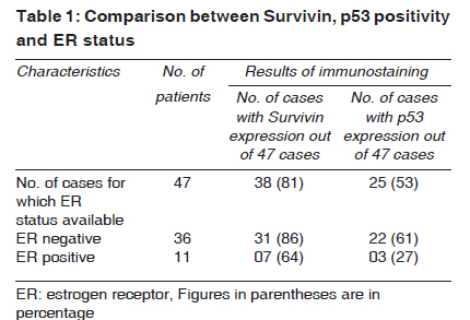
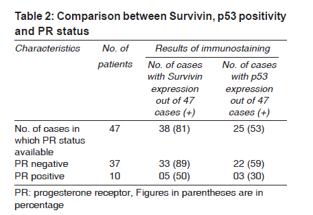
Indian Journal of Medical Sciences, Vol. 63, No. 11, November, 2009, pp. 481-490 Original Article Expression of survivin and p53 proteins and their correlation with hormone receptor status in Indian breast cancer patients Ranade KJ, Nerurkar AV, Phulpagar MD, Shirsat NV Department of Biochemistry, T. N. Medical College, B. Y. L. Nair Hospital, Mumbai Code Number: ms09090 PMID: 20075549 DOI: 10.4103/0019-5359.58877 Abstract Background : In invasive ductal carcinoma (IDC), many antiapoptotic and proapoptotic genes regulate disease outcome. Hormone receptor-mediated mechanisms have also been shown to prevent apoptosis. Therefore, relations between hormone receptor status and other molecular markers need further examination. Aims : In the present study, we analyzed the expression of apoptosis-regulating genes, viz., Survivin and mutant p53, in benign breast disease (fibroadenoma) and IDC patients. Results were then correlated with hormone receptor status of the patients. Material and Methods : Paraffin-embedded tissue samples from 63 untreated female patients with IDC and 32 female patients with fibroadenoma were used. Expression of Survivin and mutant p53 was evaluated using immunohistochemical staining method. Statistical Analysis : Fisher exact test and nonparametric correlation test (Spearman rank correlation test) were performed. Results : In fibroadenoma, 53% of patients expressed Survivin and 13% of patients expressed p53 protein. Statistically significant increase in Survivin and p53 protein expression was observed in carcinoma cases. Survivin expression correlated negatively with progesterone receptor (PR) status, but its expression was independent of estrogen receptor (ER) status. p53 expression showed negative correlation with both ER and PR status. Conclusions : Increased expression of Survivin and p53 in IDC patients and correlation with hormone receptors suggest that Survivin and p53 along with hormone receptors status are likely to contribute significantly to apoptosis resistance and may serve as therapeutic target that could increase the effectiveness of conventional breast cancer therapy.Keywords: Apoptosis, fibroadenoma, invasive ductal carcinoma, p53, survivin Introduction Breast cancer is a hormone-dependent cancer, and the presence of estrogen receptor (ER) and progesterone receptor (PR) in tumors is used clinically to predict the likelihood of response to hormonal therapies. But hormone receptor expression in human breast cancer cells does not always reflect tumor response to therapy. There could be diverse gene networks and metabolic and cell-regulatory pathways through which these hormones operate to achieve their widespread effects on breast cancer cells. Therefore, relations between hormone receptor status and other molecular markers need further examination. Survivin belongs to the group of ′inhibitors of apoptosis′ (IAP) family proteins. IAP family members inhibit apoptosis by inhibiting one or more caspases. [1] Survivin also plays a role in proliferation by regulating mitosis. [2] Survivin is undergoing intensive investigations as a potential tumor marker because of the large difference in expression between normal and malignant tissues, its causal role in cancer progression and its possible involvement in tumor cell resistance to radiation and chemotherapeutic drugs. [3],[4] In one of the recent clinical studies, the clinical and immunological responses to the Survivin-derived peptide vaccine were evaluated. [5] Though many investigations are going on, Survivin expression has not been studied much in Indian breast cancer patients. p53 is the primary arbiter of the mammalian cells′ response to stress. In its normal form, it supports apoptosis and thus has a regulatory function over the cell cycle. In its mutant form, it inhibits apoptosis, loses the control on cell cycle progression and thus helps tumor formation. Alteration of p53 is the most common mutation in all types of human cancers. [6] The aberrant cellular expression of p53 is a good prognostic biomarker of the stage and malignant vigor of cancer. [7] The current study was designed to investigate expression of Survivin and mutant p53 proteins in paraffin sections of benign breast disease patients and breast carcinoma patients. The outcome of this study was then correlated with hormone receptor status of the patients. Material and Methods The present study was carried out on retrospective samples. Paraffin-embedded samples between the years 2000 and 2006 were randomly selected for this study. Histopathologically proved and graded invasive ductal carcinoma (IDC) and fibroadenoma tissues were obtained from the pathology department of our institution. Samples were obtained after being fixed in 10% buffered formalin and embedded in paraffin for immunohistochemical analysis. This work was approved by our Institutional Ethics Committee. The clinical data of each patient was maintained, inclusive of age, sex and hormone receptor status. Tissue specimens Patients with cancerous lesions: The present study included 63 untreated female patients with invasive ductal carcinoma, their ages ranging from 22 to 50 years (median age, 36 years). Histopathologically the tumors were categorized as IDC I, IDC II and IDC III (21 cases each) using Bloom Richardson′s scoring and grading. Patients with benign breast lesions: Benign breast (fibroadenoma) lesions of 32 patients, their ages ranging from 18 to 52 years (median age, 35 yrs), were studied. Inclusion/ Exclusion criteria All biopsies were collected from patients in a specified age group who had not received any therapy before surgery. Cases other than IDC and fibroadenoma were not included. Immunohistochemical analysis Tissue sections were cut at 5 µm and mounted on poly-l-lysine-coated glass slides (sigma, St. Louis, MO, USA). Antigen retrieval was then performed by heating slides immersed in citrate buffer, pH 6, in microwave oven. [8] Primary rabbit polyclonal antibodies against p53 (SC 6243) and Survivin (SC 10811) were obtained from SantaCruz Biotechnology (Santa Cruz, USA) and used at a dilution of 1:100 in 1% Bovine serum albumin (BSA). Horseradish peroxidase-linked antirabbit IgG (NA 934V) obtained from GE Healthcare UK Lt. (Buckinghamshire, UK) was used as a secondary antibody, and the bound antibody was detected using 3, 3′ diaminobenzidine as a substrate and Harris′ hematoxylin as a counterstain. Positive and negative controls were included in each batch of slides [Figure - 2]A-B. The correlation between the level of expression and the histological grade was analyzed using the Fisher exact test. Nonparametric correlation test (Spearman rank correlation test) using Spearman correlation coefficient (r) was done between expressions and hormone receptor status. Interpretation of slides: The immunohistochemical analysis of Survivin and p53 expression in benign breast lesions and invasive ductal carcinoma (IDC) of breast was carried out on the basis of the percentage of cells showing staining. The level of expression was scored as follows: 0 = negative, less than 5% of cells staining 1+ = weak staining, between 6% and 25% of cells staining 2+ = moderate staining, between 26% and 50% of cells staining 3+ = medium strong staining, between 51% and 75% of cells staining 4+ = strong staining, more than 75% of cells staining The scores of ER/PR status obtained were then correlated with Survivin and mutant p53 expression. Statistical analysis Statistical analysis was performed using Graphpad Instat 3 software. Fisher exact test was used to find out the significant difference in expression of Survivin, p53 in benign and malignant tissues. Spearman′s correlation coefficient (r) was used to find out correlation between hormone receptor status and genetic expressions. Results Survivin expression was observed in 17 (53%) out of 32 benign breast cases. Survivin was mainly immunolocalized in the cytoplasm. However, 13% (4/32) of benign cases showed distinct nuclear expression along with cytoplasmic expression [Figure - 1]A. One benign case showed only nuclear expression. The follow-up study ranging from 4 to 5 years in these patients did not show any incidence of breast cancer development. The immunohistochemical analysis of invasive ductal carcinoma of breast showed very high levels of Survivin expression in all grades. In IDC I and II cases, 90% and 86%, respectively, showed Survivin expression; whereas in IDC III, 71% of the cases showed Survivin expression. Overall, statistically significant increase in the level of Survivin expression was observed in IDC as compared to benign cases (P = 0.0035, Fisher exact test). In IDC, Survivin expression was found to be localized in cytoplasm as well as in nucleus. Weak-to-strong Survivin expression was observed in all grades of IDC [Figure - 1]B-D. The percentages of different levels of expression of Survivin in benign and IDC cases are illustrated in Graph 1A. In immunohistochemical analysis of p53 protein, it was observed that only 13% (4/32) [Figure - 1]E of the cases expressed p53 protein in benign breast cases. In IDC I, II and III, the distinct nuclear expression of p53 was seen in 33%, 52% and 67% of the cases, respectively, throughout the tumor tissue [Figure - 1]F-H. [SUPPORTING:1] Thus statistically significant increase in mutant p53 protein expression was observed in carcinoma cases as compared to benign breast cases (P = 0.003, Fisher exact test). Moreover, the intensity of the staining was also noted to be increased with increase in histological grades. The percentages of different levels of expression of p53 in benign and IDC cases are shown in Graph 1B. Out of 63 IDC cases, ER [Table - 1] and PR [Table - 2] status was obtained from 47 cases, and correlative studies between hormone receptor status and immunohistochemical staining of Survivin and p53 were done in these 47 cases only. When comparison of immunohistochemical staining of Survivin and p53 with hormone receptor status was done in 47 breast carcinoma patients, it was observed that Survivin expression correlated negatively with progesterone receptor (Spearman r = −0.3146; P = 0.0312; 95% confidence interval, −0.5580 to −0.02139), but its expression was independent of ER status of the patients. p53 expression showed negative correlation with ER status (Spearman r = −0.3417; P = 0.0188; 95% confidence interval, −0.5785 to −0.0516) and PR status (Spearman r = −0.2950; P = 0.0441; 95% confidence interval, −0.5429 to 0.0002) of the patients. Discussion IDC is found to be the most prevalent type of breast carcinoma. In the present investigations, we studied the expression of Survivin and mutant p53 in benign breast disease (fibroadenoma) and in invasive ductal carcinoma lesions by immunohistochemical staining. These findings were then related with hormone receptor status of the patients. Survivin is a member of the ′inhibitors of apoptosis′ gene family that controls mitotic progression and induces tumor cell invasion. Its overexpression has been shown to be associated with parameters of poor prognosis in most of the human cancers, including carcinoma of the lung, esophagus. [9],[10] In the present study, statistically significant difference in the expression of Survivin between benign breast disease (53%) and breast carcinoma (83%) (P = 0.0035, Fisher exact test) was observed. Our result is consistent with the observation of Nassar et al., who had reported 81% Survivin positivity in breast cancer cases; [11] whereas according to Zhang et al., Survivin was expressed in 42.7% of benign breast tumors. [12] Normally Survivin is undetectable in terminally differentiated adult tissues; therefore, Survivin expression in benign cases is likely to be the result of dysplastic transformation of the breast epithelium. As Survivin plays a dual role, as apoptotic inhibitor and as mitotic effector, its localization was of interest to the scientists. It has been shown to be present as a cytoplasmic and nuclear protein in various cancer patients. [13],[14] Recent evidence shows that the direct interaction of Survivin with the nuclear export receptor crm1 is critically involved in its intracellular localization and cancer-relevant functions. [15] Nuclear Survivin expression has been shown to be associated with poor prognosis in non-small-cell lung cancer and esophageal squamous cell cancer. [9],[10] In the present investigation, 1 benign case showed only nuclear staining of Survivin; whereas in 4 patients, both nuclear staining and cytoplasmic staining were observed. The follow-up study ranging from 4 to 5 years for these patients showed no development of breast carcinoma. Thus Survivin expression in benign breast tumor is unlikely to be indicative of progression of malignant transformation, but large number of cases are required to be studied to draw any conclusion. It was Chu et al.[16] who had demonstrated that Survivin expression increases with increased histological grades of the disease. However, in our study we observed slight decrease in expression of Survivin as the histological grade increased. In breast cancer, a hormone-dependent cancer, several factors may be involved in regulating cell proliferation and death. Survivin being an inhibitor of apoptosis, it exerts its antiapoptotic function by deregulating the caspase cascade and thus provides resistance to apoptosis. However, apoptotic activity has been shown to increase with increasing grade of malignancy. [17],[18],[19] In such scenario, as apoptotic index and apoptosis increase, there may be many proapoptotic mechanisms which might have triggered to induce apoptosis. Such up-regulated proapoptotic mechanisms may overcome Survivin′s inhibitory properties and could reduce its expression slightly. This could be one of the several mechanisms and reasons that may be responsible for reduced Survivin expression in IDC III (71%) than in IDC I (90%) and in IDC II (86%), respectively. The exact mechanism for this phenomenon is still unknown; and since the number of samples studied is less, further evaluation is needed. In the present study, out of the total number of IDC cases studied, 6% (4/63) showed only nuclear expression of Survivin and 27% (17/63) showed nuclear and cytoplasmic expression, whereas 49% (31/63) of cases showed only cytoplasmic expression. The follow-up study ranging from 2 to 5 years in 10 patients with nuclear expression of Survivin observed about 50% survival rate. Steroid receptor analysis is the only widely accepted prognostic/ predictive marker in breast cancer treatment. When Survivin expression was correlated with hormone receptor status of the patients, it was observed that Survivin showed statistically significant negative correlation with PR (Spearman r = −0.3146; P = 0.0312; 95% confidence interval, −0.5580 to −0.02139), but its expression was independent of estrogen receptor status. Similar observations were obtained by Chu et al.,[16] who stated that Survivin is independent of ER status of the patient; whereas Span et al.[20] have shown negative correlation of PR status with Survivin expression in breast carcinoma. Ryan et al. observed that increased Survivin levels were significantly associated with negative hormone receptor status. [21] It has been demonstrated that progesterone at relatively high physiological concentrations, but comparable to those seen in plasma during the third trimester of human pregnancy, exhibited a strong antiproliferative effect on breast cancer cells and induced apoptosis. [22] Inversely, Survivin inhibits apoptosis and promotes cell proliferation. This may be the reason for negative correlation obtained between PR status and Survivin expression in most of the IDC patients Disruption of p53 gene function seems to have a pivotal role in carcinogenesis. It has been demonstrated by Rohan et al.[23] that p53 gene changes occur before the development of breast cancer and therefore influence breast cancer risk. p53 has also been reported to regulate Survivin expression. [24] In our study, 13% (4/32) of benign cases showed nuclear p53 expression. Sirotkovic et al.[25] demonstrated 19% of benign cases with nuclear p53 expression. The follow-up study in these 4 benign patients showed no development of breast cancer for at least 4 to 5 years. These findings are contradictory to the findings by Rohan et al., who concluded that p53 immunopositivity detected in normal or benign tissue is associated with increased risk of subsequent breast cancer. But to draw any conclusion, follow-up study of a large number of cases needs to be carried out. Statistically significant (P = 0.003, Fisher exact test) increase in mutant p53 expression was observed in IDC cases studied as compared to benign cases. The expression was found to increase with increasing grades. p53 was expressed in 33% of IDC I cases, while 52% and 67% of cases of IDC II and IDC III, respectively, were positive for p53. The increased p53 expression with grades and stages of various malignancies was demonstrated by many workers. [26],[27] A statistically significant negative correlation was observed between mutant p53 expression and ER status (Spearman r = −0.3417; P = 0.0188; 95% confidence interval, −0.5785 to −0.0516) and PR status (Spearman r = −0.2950; P = 0.0441; 95% confidence interval, −0.5429 to 0.0002) of the patients. Similar results were obtained by Putti et al.[28] and Pinero-Madrona et al.[29] p53 overexpression was also observed in patients with negative hormone receptor status by Nishimura et al.[30] Recently one study stated that proportion of ER-negative cases was higher than ER-positive cases in south India. [31] This shows that there could be several etiological factors specific to Indian conditions, which may be regulating hormone receptor status and indirectly influencing genetic expression. As ER-negative breast cancers have poor prognosis, the assessment of various other molecular markers and more correlative studies between hormone receptor status and various genetic expressions need to be carried out in the Indian population to understand the biological behavior of such tumors, which may provide an approach for study and use of newer promising agents in treatment of ER-negative breast cancer. Summarizing our observations, we state that increased expression of Survivin and mutant p53 in IDC patients compared to benign cases is likely to contribute significantly to apoptosis resistance. Survivin and p53 expression and their correlative studies with hormone receptors status may also serve as therapeutic target. Though Survivin is likely to contribute to apoptosis resistance, its role in predicting prognosis is still unclear. A study with large sample size, along with clinical follow-up data, is required to check whether Survivin and p53 can serve as prognostic markers and whether they can be used as target along with ER, PR status of the patient to decide on more optimal treatment modalities, which will increase the effectiveness of conventional breast cancer therapy. Acknowledgments We sincerely thank Mr. Umesh Kadam, technician, Advanced Centre for Treatment, Research and Education in Cancer, for the technical help provided by him. References
Copyright 2009 - Indian Journal of Medical Sciences The following images related to this document are available:Photo images[ms09090f2.jpg] [ms09090f1.jpg] [ms09090t2.jpg] [ms09090g1.jpg] [ms09090t1.jpg] |
| |||||||||
