 |
| About Bioline | All Journals | Testimonials | Membership | News |
|
||||||
|
||||||
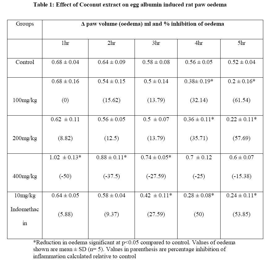
African Journal of Food, Agriculture, Nutrition and Development, Vol. 10, No. 10, 2010 pp.4286-4300 Article ANTI-INFLAMMATORY AND ANTI-ULCEROGENIC EFFECT OF ETHANOL EXTRACT OF COCONUT (COCOS NUCIFERA) ON EXPERIMENTAL RATS Anosike CA*1 and O Obidoa2 1Department of Biochemistry, University of Nigeria, Nsukka, Enugu State, Nigeria. Code Number: nd10112 ABSTRACT The effect of the ethanol extract of coconut on egg albumin- induced inflammation in rat hind paw, hypotonicity induced haemolysis of human red blood cells and indomethacin – induced gastric ulcer in Wistar rats, was studied. Fifty adult rats of either sex of weight 120-200g were divided into ten experimental groups of five rats each; five groups were used for the inflammation test, while the other five groups were used for ulcer test. Inflammation was induced by injecting 0.1ml undiluted fresh egg albumin (philogistic agent) into the subplantar surface of the right hind paw of the rats. Ethanol extract of coconut with doses of 100, 200 and 400mg/kg, and indomethacin (100mg/kg) were administered intraperitoneally to separate groups of the rats one hour before inducing inflammation. The control group received equivalent volume of normal saline (vehicle). Ulcer was induced in the rats by the administration of indomethacin (50mg/kg) (p.o.) using standard procedures. Coconut extract with doses of 100, 200 and 400mg/kg, and ranitidine (100mg/kg) were administered orally to separate groups of the rats thirty minutes before inducing ulcer. The control group received equivalent volume of normal saline (vehicle). The percentage ulcer inhibition was taken as the measure of the protection against ulcer offered by the coconut extract. The effect of the coconut extract on haemolysis induced by distilled water was evaluated by incubating various concentrations of the extract with red blood cells and distilled water. The effect of the standard anti-inflammatory drug, indomethacin was determined as a positive control. Changes in absorbance were used to assess the extent of haemolysis, hence membrane stabilization. From the results obtained, rats treated with 100 and 200mg/kg of the extract showed significant reduction of oedema at the later phase of inflammation and also reduced the ulcer induced by indomethacin, with 100mg/kg and 200mg/kg doses having an ulcer inhibition of 65.4% and 67.9% respectively; 400mg/kg of the extract increased the paw oedema of the animals and also evinced an increase in ulceration when compared to control. The coconut extract gave a dose dependent reduction in the haemolysis induced by distilled water. This suggests that the extract at low doses has potential anti-inflammatory and anti-ulcerogenic effect. Key words: Coconut extract, ulcer, inflammation, haemolysis INTRODUCTION The coconut palm (Cocus nucifera L.) locally referred to as kwa kwa in Hausa, Aku oyibo in Ibo, Agbon in Yoruba and Isip oyon in Efik, is a member of the family Arecaceae (palm family). It is the only specie of the genus cocos which grows throughout the tropical countries including Nigeria. The coconut palm is large, growing as tall as 30 meters, with pinnate leaves 4-6m long. Every part of coconut is useful and medicinal to mankind, hence it is known as the “Tree of life” [1, 2]. In traditional medicine around the world, the coconut is used to treat a wide variety of health problems. Its water (coconut water) is used as an intravenous fluid to counteract the effects of drug overdose, poisoning and adverse drug reactions [1, 3]. Coconut water is also a source of quick energy, boosts energy and endurance. It is used in place of dextrose\glucose in medical emergencies. During World War II, young coconut water was used as an emergency room glucose supply in the absence of sterile glucose [1]. Coconut water also relieves the symptoms associated with Crohn’s disease, an illness in which the intestines are infected [4]. Indigenous people of tropical countries use young coconut juice in the treatment of stomach upsets, diarrhoea and dysentery, ulcerative colitis and stomach ulcers [5]. The most abundant nutrient in the coconut is fat, which makes up more than a third of its mature weight. The lauric acid content of coconut endows it with antimicrobial properties. As such, coconut is useful in the treatment of digestive tract infections [6]. It is also used to expel intestinal parasites like tapeworms and Helicobacter pylori, which are responsible for indigestion and ulcer [7]. The oil can be processed and extracted as an organic product which, can be employed in the cosmetic industry for skin care to moisturize the skin, relieve dryness, flaking and prevent stretch marks. It is used for wounds, bruises, burns, rashes, eczema, and dermatitis. It also supports the natural chemical balance of the skin and provides protection from the damaging effects of ultraviolet radiation from the sun [7]. Coconut, due to its contents of caprylic acid, which is fungicidal, is used in the treatment of fungal skin infections such as athlete’s foot, thrush, ringworm and candidiasis [8]. In modern medicine, coconut is used as an immune system boaster in infants [6]. It improves digestion and absorption of other nutrients such as vitamins, minerals and amino acids; prevents obesity and overweight problems by increasing metabolic rate, regulates thyroid function, boosts energy and fights fatigue [9]. Coconut is used in the treatment of mal-absorption of fat such as cystic fibrosis and enteritis. It improves insulin secretion and enhances the utilization of blood glucose. This forms the basis for its use in the management of diabetes [10]. Coconut is effective in the treatment and prevention of heart disease, chronic fatigue syndrome, osteoporosis, gall bladder disease, Crohn’s disease, prostate enlargement and cancer because of its composition of high and medium chain fatty acid [7]. It also reduces inflammation and allergic reactions due to its anti-histamic effect [11]. The coconut palm has a multitude of industrial uses. It provides raw materials for industries such as the wood, furniture and food industry. Its products include: timber, food, fermented and unfermented drink, alcohol, thatching material, splint fibres for making baskets, masts rope, hats, brushes and broom. The palm leaves are used as a source of fuel and shelter. It produces utensils for household use such as cups, bowls; oils for food, illumination, soap and margarine production and ointment [12]. The residue after extraction is used in feeding domestic animals and as fertilizer. Several food uses also exist for coconut products. Coconut is highly nutritious; rich in fibre, vitamins, and minerals. It is classified as a “functional food” because it provides many health benefits beyond its nutritional content [2]. This work was aimed at investigating the effect of the ethanol extract of coconut on egg albumin induced inflammation and gastric mucosal damage induced by indomethacin on Wistar albino rats, and the membrane stabilization ability of the extract on hypotonicity induced haemolysis of red blood cells. MATERIALS AND METHODS Animals – Fifty adult Wistar rats of either sex of weight 120-200g obtained from the animal house of the Faculty of Biological Sciences, University of Nigeria, Nsukka were used for the study. They were divided into ten experimental groups of five rats each, housed in separate cages and acclimatised for seven days before the experiment. They were maintained ad libitum on water and growers mash (Pfizer Feeds, Aba) bought from Nsukka market. The research was conducted in accordance with the ethical rules on animal experimentation approved by the ethical committees of the Faculty of Biological Sciences, University of Nigeria, Nsukka. Plant Materials Matured coconut seeds purchased from the Nsukka local market was cracked and the fresh nuts chopped into tiny bits and sundried. The dried coconut was ground with a mechanical grinder and macerated in absolute ethanol for 24hrs and filtered with a white cotton cloth. The filtrate was concentrated to dry paste using a rotary evaporator at an optimum temperature of 40° -50° C. Preparation of Blood Sample Fresh blood samples (5ml) were collected from healthy donors into plastic tubes containing 1ml of 3.8% sodium citrate. These test tubes were centrifuged at 300rpm for 10mins. The red cell pellets were collected and re-suspended in normal saline equal to 2 times the volume of the supernatant. Acute toxicity study The acute toxicity test was carried out by a modified method of Lorke [13], to define the range of lethal dose and safe dose for the extract. Swiss albino mice were starved of food but allowed access to water prior to the study and were then grouped (three mice per group). They were treated intraperitoneally with different doses of the extract (50, 100, 400, 600, 1000, and 1500mg/kg). The animals were then observed for 24hrs for nervousness, dullness, in-coordination or death. Anti-inflammatory test The anti-inflammatory test was carried out using a philogistic agent - induced rat hind paw oedema as a model for acute inflammation [14]. The philogistic agent employed in this study was fresh egg albumin [15]. Twenty five (25) adult wistar rats of either sex (120g – 200g) were divided into five experimental groups of five rats each. They were fasted and deprived of water for 18hrs before the experiment. Deprivation of water was to ensure uniform hydration and to minimize variability in oedematous response [16]. Various extract doses (100, 200 and 400mg/kg) suspended in normal saline were administered intraperitoneally into groups I, II and III of the rats. Control group received equivalent amount of normal saline and the reference group was administered 100mg/kg indomethacin. One hour post treatment, inflammation of the hind paw was induced by injecting 0.1ml of undiluted fresh egg albumin (philogistic agent) into the subplantar surface of the right hind paw. This treatment was found to cause swelling of the paw which retained about the same degree of oedema for 3 hours. The right hind paw volumes of the rats were taken on the principle of volume displacement using LETICA Digital Plethysmometer (LE 7500) immediately before the experiment (zero time) and at 1hr intervals after the injection of egg albumin for a period of 5hrs. Average oedema at every interval was assessed in terms of difference in volume displacement after injecting the philogistic agent and zero time volume displacement of the injected paw (Vt – V0 ). Percent inhibition of oedema was also calculated for each dose [17], using the relation;
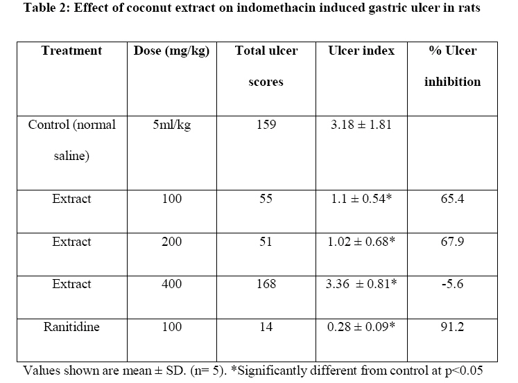
Where a = mean paw volume of treated rats after egg albumin injection Indomethacin induced ulcer This assay was carried out using a standard method [19]. Twenty five adult rats randomly divided into 5 groups of 5 rats each were deprived of food for 18hrs and treated per orally with normal saline and varying doses of the plant extract. The extracts and drugs used were freshly prepared as a suspension in normal saline and administered orally to the animals in 5ml/kg doses. Group 1 (normal control) was treated with normal saline (5ml/kg), while groups II, III and IV were treated with 100mg/kg, 200mg/kg and 400mg/kg of the coconut extract respectively. Group V (reference group) was administered 100mg/kg of ranitidine (standard anti ulcer drug). Thirty minutes later, 50mg/kg of indomethacin was administered (p.o) to the rats. After 8hrs, each animal in the groups was anaesthetised to unconsciousness using chloroform and the stomach removed and opened along the greater curvature, pinned flat on a board, examined and scored for ulcer. Erosions formed on the glandular portions of the stomach were counted and the ulcer index calculated as described by Main and Whittle [20]. The ulcer was counted and scored as 0 = no ulcer; 1 = superficial ulcer; 2 = deep ulcer and 3 = perforations. The sum of all the lesions/ulcer in all the animals for each group (total ulcer score) was used to calculate the ulcer index. Percent ulcer inhibition was calculated relative to control. Determination of membrane stabilization effect of coconut extract on hypotonicity induced haemolysis of red blood cells The effect of the coconut extract on haemolysis induced by distilled water was evaluated by incubating various concentrations of the extract with red blood cells and distilled water. This assay was carried out by a modified method of Shinde [21]. The effect of the ethanol extract of coconut on haemolysis induced by distilled water was evaluated by incubating 0.1ml solution containing graded concentrations (mg/ml) of the extract with 0.1ml of the sodium citrate treated blood, 1.8ml of normal saline and 0.5ml of distilled water in a test tube at 37° C in a water bath for 1hr. After the incubation, the test tubes were centrifuged at 300rpm for 10mins. The absorbance of the supernatants collected was read at 418nm. These experiments were done in triplicates and mean absorbance values taken. The effect of the standard anti-inflammatory drug, indomethacin was determined as a positive control. Changes in absorbance were used to assess the extent of haemolysis; hence membrane stabilization. Percentage inhibition of haemolysis by the extract was calculated thus:
Where OD1 = Absorbance of isotonic solution Statistical analysis: This was done using SPSS version 14.0 (SPSS Inc. Chicago, IL.USA). All values are expressed as mean ± SD. Data were analysed by one-way ANOVA and difference between means was assessed by a two-tailed Student’s T-test. P<0.05 was considered statistically significant. RESULTS The LD50 of the crude ethanol extract of coconut was calculated to be 1000mg/kg. All the doses used in this study were therefore carefully chosen to exclude the lethal range. The data showed that fresh egg albumin induced oedema in the rat paw which was sustained over a period of the 5hrs (Table 1). Groups treated with 100mg/kg and 200mg/kg of coconut and 100mg/kg indomethacin showed a significant reduction in paw oedema (p<0.05) at the later phase of inflammation when compared to control, while the group treated with 400mg/kg of coconut registered increased paw oedema. The increase was significantly higher (p<0.05) than that of control and the other groups (Table 1). Indomethacin was found to induce gastric ulcer in all the experimental groups (Table 2). Groups administered with 100mg/kg and 200mg/kg of coconut had significant reductions (p<0.05) in the ulcer indices as compared to control, thus showing a high percentage level of ulcer inhibition, 65.4% and 67.9% respectively, though not as high as that of the standard drug, ranitidine. The group treated with 400mg/kg of coconut, on the contrary, showed an increase in ulceration as is shown by its high ulcer index, which is higher than that of control. The coconut extract showed a dose dependent significant reduction in the haemolysis of the red cells induced by distilled water (Table 3). DISCUSSION Inflammation is a complex biological response of vascular tissues to harmful stimuli, such as pathogens, damaged cells or irritants. It is also a localized protective response elicited by injury which serves to destroy both the injurious agent and the injured tissue [22]. Inflammation is classified as acute or chronic depending on the type and duration of the antigen challenge and is mediated by some chemical substances such as histamine, serotonin, Slow Reacting Substances of Anaphylaxis (SRS-A) (leukotrienes), prostaglandin and some plasma enzyme systems such as the complement system, the clotting system, the fibrinolytic system and kinin system. These agents can originate locally or from cells that infiltrate the site of insult [23]. Oedematous response in inflammation occurs due to the action of inflammatory mediators such as histamine, serotonin and bradykinin at the site of a local inflammatory insult [24]. These mediators cause vasodilatation and increased permeability of blood vessels leading to the exudation of plasma proteins and fluids into the tissues. The early phase of oedema, beginning from 1hr after the administration of the irritant, is due to the release of histamine and serotonin, while the later phase, occurring from 3 to 5hrs after the administration of the irritant is induced by bradykinin, protease, prostaglandin and lysosome [24, 25]. In this study, oedema inhibition was observed with the lower doses of the coconut extract, and was more pronounced in the 2nd phase of inflammation post administration of extract. This activity may be due to the suppression of the prostaglandin and kinin formation induced by the philogistic agent. Oedema inhibition in the 1st phase of inflammation post administration suggests an inhibitory effect on histamine and serotonin secretion. High dose- (400mg/kg) of the extract- was pro-inflammatory, rather than anti-inflammatory. This result is paradoxical, since previous work [11] reported that coconut has good anti-inflammatory activity. This study suggests that the extract could be harmful at high doses. In the present study, significant protection against indomethacin induced gastric mucosal damage was observed in the animals treated with 100mg/kg and 200mg/kg of the coconut extract. This suggests that the extract is anti-ulcerogenic only at low doses. An earlier study [7] showed that coconut oil could be used in the treatment of stomach ulcers. Stomach ulcer results from an injury or damage to the gastric mucosal lining of the stomach which could be a result of excess or an overproduction of hydrochloric acid, an acid normally present in the digestive juices of the stomach or due to complications resulting from an infection with Helicobacter pylori [26]. It also results due to excess intake of non- steroidal anti-inflammatory drug (NSAIDs) such as indomethacin, aspirin and ibuprofen. Gastric ulcers induced by these drugs is related with the inhibition of cyclooxgenase 2 enzyme which synthesizes prostaglandin needed to maintain the integrity of the gastric lining of the stomach [25]. In this study, the ethanol extract of coconut at lower doses exhibited anti-ulcerogenic effect against indomethacin induced gastric ulcer, which is comparable with that obtained for ranitidine, an anti-acid used to neutralize intraluminal acid, improve gastric microcirculation and reduce the absorption and concomitant adverse drug interactions of many NSAIDs [27]. This result is in agreement with that of Nneli and Woyike [28], which showed that coconut milk and water exhibited anti-ulcerogenic effect against indomethacin induced ulcer and suggests that coconut may act by reducing the intestinal absorption of indomethacin and its resulting drug interactions. The test group treated with 400mg/kg of the extract showed an increase in ulceration. This result is similar to that obtained from the egg albumin- induced inflammatory test, in which the group treated with 400mg/kg of the extract showed increased paw oedema compared to that of the control group, suggesting that coconut could be deleterious at high doses. The coconut extract exhibited high membrane stabilization effect against hypotonicity induced haemolysis of the red cells as is shown by the percent inhibition of haemolysis. This inhibition of haemolysis was found to be dose dependent, increasing with increased concentration of the extract in the medium and was comparable with that of indomethacin, a standard anti-inflammatory drug. Hypotonicity induced haemolysis of red blood cells occurs due to water uptake by the cells and leads to the release of haemoglobin which absorbs maximally at 418nm. Hence, the reduced optical density at 418nm obtained for the various coconut test samples is a reflection of the stabilization of the red cell membrane caused by the extract. During inflammation, there are lyses of lysosomes which release their component enzymes which produce a variety of disorders [29]. Since human red blood cell (RBC) membranes are similar to lysososmal membrane, human RBC stabilization was therefore used as a method to study the mechanism of action of anti-inflammatory agents [30]. A previous study reported that coconut reduces inflammation and allergic reactions due to its anti-histamic effect [11]. The stabilizing effect of coconut extract on lysosomal membranes as seen in this study suggests a possible mechanism of action for the anti-inflammatory effect of coconut. Results from this study have shown that coconut has potential as an anti-inflammatory and anti-ulcerogenic agent, but only at low doses, high doses could be pro-inflammatory and ulcerogenic. ACKNOWLEDGEMENT We acknowledge the assistance of the technical staff of the Department of Biochemistry, University of Nigeria, Nsukka. REFERENCES
Copyright 2010 - African Journal of Food Agriculture, Nutrition and Development The following images related to this document are available:Photo images[nd10112t1.jpg] [nd10112t3.jpg] [nd10112t2.jpg] |
| |||||||||

