 |
| About Bioline | All Journals | Testimonials | Membership | News |
|
||||||
|
||||||
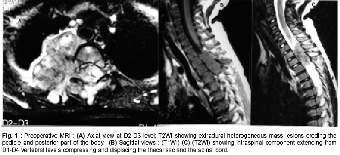
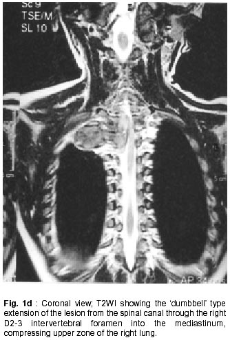
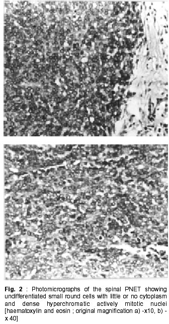
Neurology India, Vol. 50, No. 1, March, 2002, pp. 75-80 CASE REPORT Primary Intraspinal Primitive Neuroectodermal Tumor (PNET) : A Rare Occurrence M.J.Virani, S. Jain Department
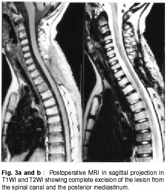
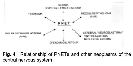
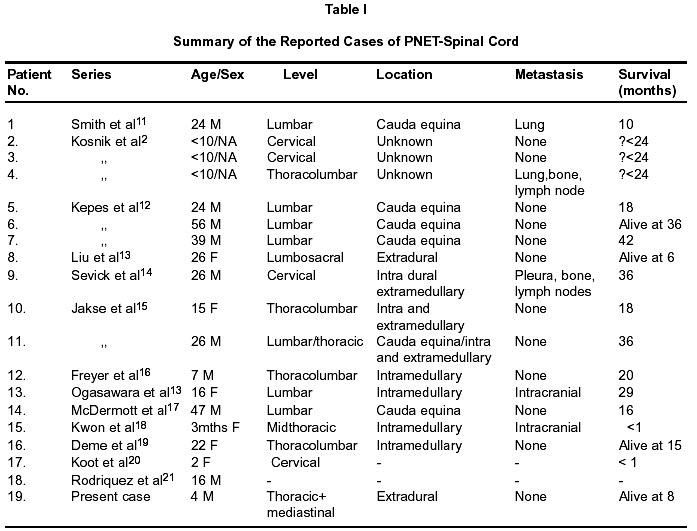
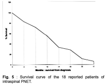
of Neurosurgery, Jaslok Hospital and Research Centre, Mumbai - 400 026, India. Accepted for publication : 18th December, 2000. Code Number: ni02018 Summary The concept of primitive neuroectodermal tumors (PNETs) has been evolving for many years, as has been its nomenclature. A 5 year old boy presented with pain in lower cervicodorsal region and left leg. Preoperative MRI of the spine and paravertebral region revealed a hyperintense lobulated lesion extending from D1-D4 with a large intraspinal and thoracic component. A total removal of tumor was achieved via a dorsal laminectomy and right posterolateral thoracotomy. The pathological findings were consistent with PNET. Post operative neurological examination had been unremarkable. Six months follow up scan showed no recurrence. A review of the literature shows that only 18 cases of primary intraspinal PNETs have been reported to date and the present case is exclusive, in which the tumor was thoracic, extradural in location and the child is alive at 8 months of follow up, with no evidence of tumor recurrence/metastasis. Primary intraspinal PNETs are rare tumors and carry a poor prognosis. Newer modalities of treatment should be tried to improve survival. Key words : Spinal cord, Primitive neuroectodermal tumor (PNET), Radiation, Chemotherapy, Immunotherapy. Introduction Primary neuroectodermal tumor (PNET), a term proposed by Hart and Earle defines a group of malignant neoplasms of presumed neural crest origin.1 Cases of PNET have been increasingly reported in recent years but there are still very few reports of PNET originating in the spinal cord. To date, only 18 cases of primary intraspinal PNETs have been reported in the literature. The clinical and pathological features of PNET, its management, and perspectives for the future, with reference to a case of PNET of the spinal cord, are discussed. Case Report A previously healthy 5 year old male child presented with complaints of lower cervico-dorsal pain, breathlessness on exertion, pain in left leg, lassitude, lethargy of one month duration. Family history was unremarkable. There was no history of sphincteric disturbances, weight loss and gait disturbances. On examination, vital parameters were normal. Minimal thoracic scoliosis to right was observed. Neurological examination was unremarkable except for the unsteady gait and tendency to sway to right side. Finger-nose and toe-heel tests were normal. Radiographic investigations : X-ray chest revealed a heterogeneous soft tissue shadow in right upper zone and right paraspinal region with a smooth convex borders with apparent widening of right 3rd intercostal space posteriorly. Pneumonic consolidation or posterior mediastinal mass were considered as possibilities. MRI of the spine and paravertebral region revealed a hyperintense lobulated lesion in paravertebral region extending from D1-D4 with a large intraspinal and thoracic component. Spinal cord was pushed completely to the left by the lesion (Fig. 1a-c, d). Operation : In view of MRI appearance of the tumor hemilaminectomy from C7 to D1 on right side was performed along with right posterolateral thoracotomy. The tumor was extradural in origin and was compressing the cord. Following surgery intercostal tube was inserted to prevent pneumothorax. Postsurgery good chest expansion was noted. Gross total removal of the tumor was performed under the operating microscope. Pathological findings : Tissue specimen represented a focally necrotic undifferentiated malignant tumor consisting of small round cells exhibiting little or no cytoplasm and rounded smooth contoured vesicular or dense hyperchromatic actively mitotic nuclei. Cells were arranged in compact pattern. No well-defined Homer-Wright or ependymal rosettes were noted (Fig. 2). Immunohistochemistry was performed using the primary antibodies : synoptophysin, (Dako corp.), LCA and Cytokeratin. Tumor cells showed granular immunostaining in the cytoplasmic rim with synoptophysin. Cytokeratin, LCA were absent in the tumor. Electron microscopy confirmed the presence of poorly differentiated small round cells.The diagnosis of a PNET was arrived at after histological examination, immunohistochemistry and electron microscopy. Postoperative course : Postoperative course had been uneventful. CT scan brain was normal. Post surgery neurological status showed a slightly weak right grip, unsteady gait which improved with gait training and physiotherapy. A thoracic chin occiput brace was provided. Following surgical recovery, radiation therapy was given in the form of involved field irradiation (IFI) for a period of 8 weeks. MRI at the conclusion of radiotherapy failed to reveal any recurrence or metastasis (Fig. 3). Currently, eight months after surgery he was asymptomatic, walked unsupported, and had no neurological deficits. Discussion PNETs are rapidly growing tumors with a brief duration of symptoms and a rapidly progressive course.1 The tumors encountered are difficult to classify.2 It was first described as a tumor arising in peripheral nerve, and was called neuroepithelioma.3 Hart and Earle first introduced the term primitive neuroectodermal tumor in 1973 to describe predominantly undifferentiated tumors of the cerebrum (with 90-95% of the cells being undifferentiated) that did not fulfill the diagnostic criteria for neuroblastoma, ependymoblastoma, polar spongioblastoma, medulloepithellioma or pineal parenchyma tumors. All neoplasm showing primitive poorly differentiated neuroepithelial cellls can be called primitive neuroectodermal tumors, regardless of location or cell type.2,5 Relationship of PNETs and other central nervous system neoplasms is shown in Fig. 4. In 1983, Rorke6 and Becker and Hinton7 independently reviewed this concept and published separate articles advocating that all central nervous system tumors predominantly composed of primitive neuroepithelial cells be called PNETs. They then further subclassified these tumors based on differentiation. This concept has been widely accepted, although it is still controversial. The most recent classification by world health organization tries to avoid this controversy by grouping these tumors under the category of 'embryonal tumors' with PNET used as a generic term for cerebellar medulloblastomas.8 PNETs most commonly occur in the cerebellum (medulloblastoms) but can arise in the pineal gland, cerebrum, spinal cord brain stem, and peripheral nerves.9 Primitive neuroectodermal tumors frequently metastasize via the CSF pathways to the spinal and cranial subarachnoid spaces and are highly malignant both histologically and clinically.10 Standard therapy for the PNET currently consists of gross total resection followed by craniospinal irradiation. Radiation is associated with a higher incidence of intellectual impairment endocrinological disturbances, and growth retardation in young children and results in 5 year survival rates of only 40% to 60%. Chemotherapy is the sole form of therapy used in children under two years of age, because of severe side effects of irradiation in this age group.10 Because surgery, irradiation and chemotherapy do not adequately treat PNET additional treatment modalities need to be explored.. The 18 previously reported cases along with present case are summarized in Table I. In most of these cases, efforts were made to exclude primary intracranial lesions either by imaging and/or autopsy. In present case, the extradural location makes it unlikely that this is a drop metastasis. Morever, CT scan of head failed to reveal any intracranial tumor. This case therefore appears to be exclusive and represents a primary thoracic spinal cord PNET, as neither cerebral nor cerebellar intra-axial lesion, nor peripheral neuroblatomas were seen. A review of literature shows that primary intraspinal PNETs may arise at all levels of the spine and can be intramedullary, intra - and extramedullary, extramedullary or extradural. It has been postulated that PNETs arise from neoplastic transformation of primitive neuroepithelial cells in subependymal zones.6 The clinical characteristics of spinal PNETs in the cases described so far including ours (Table I) appear to be:- i) more common in adults rather than children. 12 out of 19 cases being adults, ii) males were predominnantly affected, iii) some of the reported cases had metastasis outside neuraxis with the most frequent sites being lung, bones and lymphnodes, a tendency shared by intracranial PNETs,2,11,14 iv) most of the patients were treated with a combination of surgery, radiotherapy and chemotherapy, but despite treatment most patients did not do well, v) extremely short duration of symptoms favour rapidly growing nature of these tumors, vi) the aggressive nature of the tumor is evidenced by rapid recurrence of the tumor in most of the reported patients. The cause of death in these patients included pneumonia,12 metastatic disease,14 aggressive local spread of the diseased,18 and progressive spinal cord involvement.16 vii) the tumor was frequently located at lower spinal levels : cervical in four cases, thoracic in two, thoracolumbar in four, lumbar/lumbosacral in 7 cases. viii) as expected in rapidly growing tumors such as these, the survival is less than 2 years. Less than 40% of these patients were alive 2 year after diagnosis, about 10% at 3 year (Fig. 5). Therefore, need for newer therapeutic modality to improve the survival in these cases. PET with '8F-fluoro-2-deoxy-glucose (FDG) is an effective imaging modality for evaluating suspected tumor recurrence. Use of FDG PET imaging for spinal cord neoplasms has not yet been studied, mainly due to limitations of spatial resolution. Cidis et al22 demonstrated the role of FDG PET imaging in recurrent intramedullary PNET affecting the cervical spinal cord. Adoptive immunotherapy is currently being investigated as a possible therapy. Lymphokine - activated killer cells possess several attributes that could make them useful in adoptive immunotherapy. They are highly potent against tumors, require no prior antigen exposure to express their oncolytic effect. Their recognition mechanism is able to distinguish between normal and malignant cells and thereby spare normal tissue and they express oncolytic activity against many tumour types.10 This study was under taken by Richard et al10 to determine the potential sensitivity to the tumor cells derived from PNET. The results presented in this study support an adoptive immunotherapeutic approach, consisting of intrathecal administration of IL-2 and LAK cells as an adjuvant to the treatment of PNET. This form of therapy could eradicate residual tumour without the harmful side effects that radiation or chemotherapy produce. The optimal therapy for PNET is uncertain. Early onset of chemotherapy17,23 in conjunction with radiation therapy may improve the survival time. However the prognosis of this disease is very poor and most patients develop local recurrence. As regard to new treatment strategies are concerned, role of peripheral blood stem cell transfusion (PBSCT) is suggested in chemosensitive tumors or in cases where the patient has remissions. PBSCT after remissions prevents relapse. A trial has been conducted at Hinduja hospital, Mumbai, India, where PBSCT was employed in 21 year old male with PNET of chest wall-stage-IV. More studies are required to explore the role of PBSCT in improving the survival in these patients.24 Based on this review, we conclude that future advances in the treatment of PNETs must lie with chemotherapy and immunotherapy especially for those patients presenting with disseminated disease. This, combined with early detection, tumor identification and surgical removal and aggressive neuraxis radiation, offers hope of long term and good quality survival. It is fascinating that a tumor which may be of embryonic origin can remain latent and become manifest many years later, suggesting differences in biology involving the tumor itself or the host. References
Copyright 2002 - Neurology India. Also available online at http://www.neurologyindia.com The following images related to this document are available:Photo images[ni02018f5.jpg] [ni02018f2.jpg] [ni02018t1.jpg] [ni02018f4.jpg] [ni02018f1a-c.jpg] [ni02018f3.jpg] [ni02018f1d.jpg] |
| |||||||||
