 |
| About Bioline | All Journals | Testimonials | Membership | News |
|
||||||
|
||||||
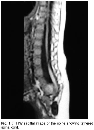
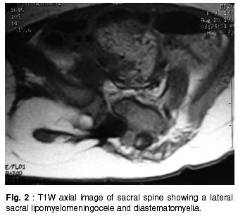
Neurology India, Vol. 50, No. 2, June, 2002, pp. 204-206 Lateral Sacral Lipomyelomeningocele : A Rare Anomaly D.S. Shetty, B.N. Lakhkar Departments of Radio Diagnosis and Imaging,
Kasturba Medical College,
Manipal, Karnataka, India. Accepted for publication : 6th June, 2001 Code Number: ni02056 Summary Lateral sacral lipomyelomeningocele is a rare spinal developmental anomaly. In the case under report, the fat attached to the neural placode was blending with the gluteal fat externally. The cord was tethered at this level. Multiple bony anomalies and diastematomyelia were associated findings. A case of lateral sacral lipomyelomeningocele with excellent imaging detail provided by the multiplanar magnetic resonance (MR) scan is reported. Key words : Lateral meningocele, Lipomyelomeningocele, MRI. Introduction Lipomyelomeningoceles are lipomas that are tightly attached to the dorsal surface of a neural placode and extend dorsally through a spina bifida to be continuous with the subcutaneous fat. The clinical presentation can be with a soft mass, sensory or motor loss or bladder dysfunction. The entity of lateral sacral lipomyelomeningocele continuous with the gluteal subcutaneous fat has not been reported in the literature. Case Report A nine month old female child weighing 6.5 kg presented with swelling over the right buttock region since birth. The swelling was progressively increasing in size. There were no other complaints. The child was able to sit up with support and the milestones were normal for the age. There was no history of retention of urine and the child was passing stools normally. Vaccination history was satisfactory. On examination, there was no jaundice, clubbing etc. Pulse and blood pressure were normal. Local examination of the gluteal region revealed diffuse swelling with no redness over it. Midline was normal, with no sinus or swelling. CNS examination revealed sluggish knee and ankle reflexes on the right side. The reflexes were normal on the left lower limb. There were no meningeal or cerebellar signs. A clinical diagnosis of hematoma or meningocele was considered and further investigations were requested. Plain X-ray of lumbosacral spine showed multiple sacral bone anomalies. Hemivertebrae and fusion anomalies of posterior elements were evident. MRI revealed tethering of the spinal cord (Fig 1). Axial images of the sacral region revealed diastematomyelia with a lateral sacral lipomyelomeningocele with right half of the cord tethered into it (Fig 2). The fat of lipomyelomeningocele was continuous with the gluteal fat. The above findings were confirmed on surgery and the tethering was released from the right gluteal region. Discussion A meningocele is an out pouching of leptomeninges through a developmental defect in the dura. The arches of the vertebrae at one or more levels are involved with protruded meningeal sac covered with only a layer of skin. The presence of spinal cord or cauda equina within the meningocele is termed myelomeningocele. This deformity mostly occurs at the stage of neuralation that prevents the neural groove from closing dorsally. One theory suggests that excessive overgrowth of neural tissue in the region of the spina bifida prevents the neural groove from fusing.1 Anterior sacral meningoceles are anomalies characterized by a focal erosion or hypogenesis of segments of the sacrum and coccyx with herniation of a CSF filled meningeal sac through the defect into the pelvis. They account for around 5% of retrorectal tumors and both sexes are equally affected.2 Lateral meningoceles have been described in the thoracic and lumbar region. They are extensions of dura and arachnoid through an enlarged neural foramen. They commonly present during fourth and fifth decade of life. Neurofibromatosis is present in approximately 85% of patients with lateral thoracic meningoceles.3 The position of the cord with respect to the meningocele sac is variable. They often occur in a setting of Marfan's syndrome or neurofibromatosis but may also be seen as isolated anomalies. Lipomyelomeningoceles are lipomas that are tightly attached to the dorsal surface of a neural placode and extend dorsally through a spina bifida to be continuous with the subcutaneous fat.4 These lesions constitute approximately 20% of skin covered lumbosacral masses.5 Lipomyelomeningoceles usually occur in the lumbosacral region of the cord and tether the cord at the level. The dorsal surface of the placode, adjacent to the lipoma, has no ependymal lining and is covered by a relatively thick layer of connective tissue mixed with islands of glial and smooth muscle fibres.6 The lipoma lies immediately external to the connective tissue in the extradural space. Patients are typically female and present before the age of six months but occasionally the condition goes undetected into childhood. The clinical presentation can be with a soft mass, sensory or motor loss or bladder dysfunction. Symptoms are progressive if untreated. In the present case, the child was nine months old and presented with a gluteal swelling. Neurological examination revealed sluggish knee and ankle reflexes on the right side. The lipomyelomeningocele was lateral in location in the sacral region, with the fat attached to the neural placode blending externally with the gluteal fat. There was associated tethering of the hemicord and diastematomyelia. Diastematomyelia is a form of spinal dysraphism characterized by midline sagittal splitting of the spinal cord of variable length. It is seen in 31% to 46% of patients with myelomeningocele.7 It is usually associated with tethering of the conus. When associated with lipomyelomeningocele, it is usually cranial to the placode. The spur, which may be bony or cartilaginous, arises from the midline dorsal surface of the vertebral body and extends dorsally. Syringohydromyelia may be found in either or both hemicords. In 15%-20% of patients with diastematomyelia, myelomeningocele of one of the hemicord is present.8 To the best of our knowledge, no association of lipomyelomeningocele with hemicord of diastematomyelia has been reported earlier. Scalloping of the pedicles, laminae and vertebral bodies adjacent to the meningocele result in an enlarged spinal canal. Butterfly vertebra and segmental anomalies of the vertebral bodies may be found in as many as 43% of affected patients. Sacral anomalies like confluent sacral foramina and partial sacral agenesis occur in upto 50% of cases.9 In the present case, the sacral vertebrae showed anomalies of hemivertebrae and fusion anomalies of posterior elements. In an individual patient, multiple anomalies are often coexisting. Therefore it is important to image the entire spine, if a cutaneous anomaly is present, which is possible with MRI. The entity of lateral sacral lipomyelomeningocele continuous with the gluteal subcutaneous fat has not been reported in the literature. Lateral meningoceles have been reported in the thoracic and lumbar regions, in association with neurofibromatosis. References
Copyright 2002 - Neurology India. Also available online at http://www.neurologyindia.com The following images related to this document are available:Photo images[ni02056f2.jpg] [ni02056f1.jpg] |
| |||||||||
