 |
| About Bioline | All Journals | Testimonials | Membership | News |
|
||||||
|
||||||
Neurology India, Vol. 50, No. 3, Sept, 2002, pp. 267-271 Human Brain : Biochemical Lateralization in Normal Subjects R. Jayasundar Department of NMR,
All India Institute of Medical Sciences,
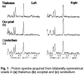
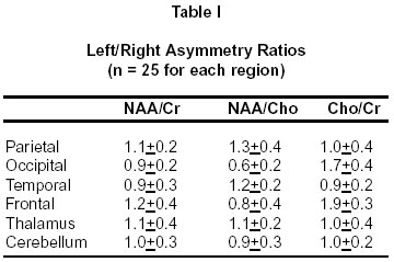
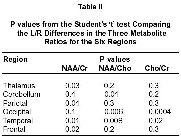
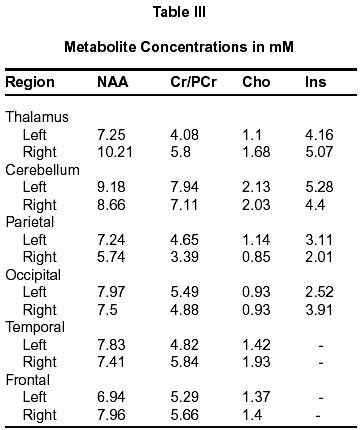
New Delhi - 110 029, India. Accepted for publication : 15th January, 2002. Code Number: ni02079 Summary Chemical asymmetries in normal human brain were studied using the non-invasive technique of volume localized proton magnetic resonance spectroscopy (MRS). The technique of STEAM was used to acquire water-suppressed proton spectra from 8 ml voxels placed in bilaterally symmetrical positions in the two hemispheres of the brain. One hundred and sixty eight right-handed male volunteers were studied for six different regions in the brain (n=28, for each region). Parietal, occipital, temporal, frontal, thalamus and cerebellum regions were studied. The focus was on metabolites such as N-acetyl aspartate (NAA), creatine/phosphocreatine (Cr/PCr) and choline (Cho) containing compounds. Ratios of the peak areas were calculated for them. Quantitation of the metabolites were carried for data on 18 volunteers. Significant interhemispheric differences in the distribution of metabolites were observed for all the regions studied. There were statistically significant differences on right and left side for the metabolite ratios in all the regions studied. The study has shown the existence of significant lateralization in the distribution of proton MR visible metabolites for all the regions studied. Key words : Magnetic resonance spectroscopy, Chemical asymmetry, Normal brain, Lateralization. Introduction The concept of cerebral dominance and hemispheric asymmetry has been a subject of lively interest since the pioneering works of Dax and Broca in the 19th century.1 A variety of techniques have since then been used to study the cerebral asymmetries and it is now well accepted that the two halves of the human brain are structurally and functionally different.2-11 In contrast to the wealth of data available on structural and functional asymmetries, there are very few studies on the invivo neurochemical asymmetries.12-14 This study presents data from 168 volunteers on biochemical lateralization in normal human brain. Material and Methods Volunteers : Normal volunteers were recruited for the study. Inclusion criteria were (i) absence of neurological deficits and (ii) right-handedness, as determined by which hand was preferred for writing. 168 male volunteers in the age group of 20 - 30 years (mean age - 25 yrs) were studied. Spectroscopy was carried out for the following regions in brain: parietal (n=28), occipital (n=28), temporal (n=28), frontal (n=28), thalamus (n=28), and cerebellum (n=28). Methods : 150 volunteers were studied at 1.5 Tesla (Siemens Helicon SP 63/84 MR scanner). The standard circularly polarized head coil was used for both imaging and spectroscopy. Images for localizing the volume of interest (VOI) were acquired in three orientations using T1W sequence ( TR msec/TE msec = 520/18). Localized water-suppressed 1H MR spectra were acquired with the STEAM RF pulse sequence.15 The experimental parameters consisted of a repetition time (TR) of 6000 msec, echo time (TE) of 135 msec, vector size of 2K data points and 128 averages.A long TR was chosen to avoid signal saturation. Bilaterally symmetrical VOIs of 8 ml each were selected from both the hemispheres. Shimming was done on the entire head as well as on the two VOIs. The entire study was carried out in one session. Care was taken to position the VOI in identical regions in all the volunteers. The spectra were processed after zero filling the original 2K data points to 4K data points. A Gaussian linebroadening of 2 Hz was applied followed by phase and baseline corrections. The focus was on the three major resonances observed in brain proton MRS, namely, N-acetyl aspartate (NAA), total Creatine (Creatine + Phosphocreatine - for brevity, this will be referred to as Cr in the rest of the text) and Choline (Cho). NAA was assigned to the peak at 2.01 ppm, Cr and Cho to those at 3.0 ppm and 3.2 ppm, respectively. The peak areas were calculated by the integration subroutine provided by the manufacturer and ratios of the peak areas, NAA/Cr, NAA/Cho and Cho/Cr were determined for each VOI. Analysis was carried out for spectra from both the hemispheres separately and this was performed in a blinded fashion to rule out the possibility of bias. The three metabolite ratios are further expressed as left (L)/right (R) asymmetry ratios, i.e., [(NAA/Cr) (left)] / [(NAA/Cr) (right)], etc. Expressing the data in this manner facilitates an estimate of interhemispheric asymmetry. In addition to the above-mentioned studies on 150 volunteers, data was acquired from 18 volunteers (3 volunteers each for the 6 different regions in the brain) at 2 Tesla using a shorter echo time (20 ms). The sequence and the other parameters used were the same except for the number of averages, which were 64. These studies were carried out and analyzed at the Biomedizinische NMR Forschungs, Max Planck Institute for Biophysical Chemistry, Goettingen, Germany. Absolute metabolite concentration in mM was determined for all the metabolites by LC Model, a user-independent fitting routine.16 Reproducibility : To assess reproducibility, five volunteers were studied four times each for a single region. The studies were done in a blinded fashion, by having the repeat volunteer included in the day's study by others without the knowledge of the researcher. Statistics : The means with standard deviation (SD) was calculated for the metabolite ratios from the different regions. The student's paired `t' test was used to compare the significance of the L/R asymmetry of these metabolite ratios. p < 0.05 was considered statistically significant. Only data from the 150 volunteers have been used for statistical analysis. Results Fig. 1 shows representative spectra from both the left and right hemispheres for some regions of the brain (thalamus, occipital and cerebellum). The L/R asymmetry ratios for the six regions studied are presented in Table I. The degree of lateralization was varied. In some individuals the asymmetry ratios were > 1.5, while in some others the L/R ratios were less than one, indicating reversal of asymmetries. The metabolite ratios for each region (from the left and right hemispheres) were also pooled separately and expressed as means+SD. P values calculated to test the significance of the differences in the metabolite ratios between the left and right hemispheres are presented in Table II. In thalamus and cerebellum, only one of the three ratios showed statistically significant L/R asymmetry. In the parietal region, the NAA/Cr showed a statistically significant asymmetric distribution, whereas NAA/Cho and Cho/Cr did not show significant L/R differences. In the occipital region, while the L/R asymmetry for NAA/Cr was not statistically significant, those for NAA/Cho and Cho/Cr were statistically significant. In the temporal region, the L/R asymmetry showed statistical significance for all the three ratios. In the frontal region, while the NAA/Cho and Cho/Cr did not show significant L/R asymmetries, NAA/Cr showed a statistically significant L/R difference. While expressing the metabolite ratios as means+SD, it was found that the large inter-individual variations made the group differences for some of the ratios in some of the regions not statistically significant. This was so for the thalamus, cerebellum and frontal regions. It is believed that the group differences have been obscured by the marked inter-individual variations. Table III presents the metabolites' concentration in mM for a single volunteer for the different regions - the data was obtained at the TE of 20 msec. The lateralization of the metabolites is varied. For example, there is considerable lateralization of NAA in thalamus (41%), parietal (21%) and frontal (15%) regions compared to that in cerebellum (6%), occipital (6%) and temporal (5%) regions. Similarly, the total Cr's lateralization is not much in cerebellum (10%), occipital (11%) and frontal (7%) compared to thalamus (42%), parietal (27%) and temporal (21%) regions. The lateralized distribution of Cho was considerable in thalamus (53%), parietal (25%) and temporal (36%) but not in the other regions (< 5%). In addition to the 3 metabolites discussed above, Inositol (Ins) also showed considerable asymmetric distribution in thalamus (22%), cerebellum (17%), parietal (35%) and occipital (55%). It is interesting to note that all the metabolites showed a consistent rightshift in thalamus and frontal regions and a left-shift in cerebellum and parietal regions. Reproducibility : The intraindividual reproducibility of the L/R asymmetry was excellent (within 5%) for all regions except the frontal lobes. This region showed more variations in the L/R ratio. But what is important is that all the regions showed consistent L/R differences on repeated studies. Discussion Extensive work has been carried out for studying the structural and functional asymmetries in human brain. In right-handed subjects, almost all the regions in the brain are known to exhibit both structural and functional asymmetries. Anatomical and Functional Asymmetry in Righthanded Subjects Frontal lobes are known to be involved in thought processes and behaviour programming.5 Structurally, the right frontal lobe has been reported to be larger than the left, and variability of the gyral pattern has been found to be greater in the left hemisphere.2,3,8 Functional asymmetries have also been reported by a number of groups.17,18 Hemispherical asymmetries at the morphological level have been reported to be commonly apparent in parietal lobes.3,8,18 The sylvian point has been reported to be higher in the right hemisphere. Associated with a lower sylvian point, the left post central gyrus, particularly its lower portion, is wider than the right. Functional asymmetry during task activation has also been reported.3,19 Temporal lobes are most extensively studied areas of the brain, because of its reported role in speech and language.6 Morphologically, the planum temporale in this region has been found to be consistently larger in the left hemisphere. Both structural as well as functional asymmetries have been reported for this region.10,11,20 The left occipital lobe is reported to be significantly wider than the right, in addition to the existence of functional asymmetry.8,18 While both structural and functional asymmetries have been reported in thalamus4,12 asymmetric blood flow to the cerebellar hemispheres in resting volunteers has been reported by Gur et al.9 In general, a greater density of cells has been reported in the left than in the right hemisphere and the left hemisphere has been found to be more extensively fissured than the right.3,6 Chemical asymmetry From the foregoing paragraphs, it is clear that there is abundant evidence from neuroanatomical and functional studies that the human brain is lateralized. In contrast, there is very little in vivo evidence of neurochemical laterality. Studies on postmortem brains have revealed chemical asymmetries in thalamus and temporal lobes. Asymmetrical distribution have been observed for glutamic acid decarboxylase, gamma-aminobutyric acid, choline acetyltransferase, and dopamine.21-24 The proton MRS studies in-vivo, in human volunteers have shown evidence of laterality in the MRS visible metabolites in the temporal and other regions in the brain.12-14 In this study, lateralization has been found for all the three major metabolites examined. Although reports on the chemical asymmetries from postmortem brains do not deal with NAA and Cr, there is strong evidence for asymmetrical distribution of ChAT in the temporal lobes and other cortical structures. Acetylcholine (ACho) is an important neurotransmitter which is considered to play a key role in intellectual activity including memory. It is synthesized from its two immediate precursors Cho and acetyl coenzymeA (acetyl CoA) by the enzyme ChAT. Cho + Acetyl CoA Û ACho + CoA An asymmetrical distribution of ChAT would result in a similar asymmetrical distribution of ACho, which is considered to contribute to the Cho resonance at 3.2 ppm.25 Correlation between MRS and other techniques Under normal physiological conditions, cerebral blood flow (CBF) is not only a direct indicator of cerebral energy metabolism, but is also closely coupled to functional activity of neurons and concomitant energy metabolism. EEG, another measure of the brain activity is also considered to be closely related to CBF and metabolism.5 A strong correlation between EEG, blood flow and cerebral metabolism using PET has been found.26-28 In addition, correlation between cerebral glycolysis as measured by PET and proton MRS has also been demonstrated.29 From the above, it can be inferred that the metabolites observed in proton MRS play an important role in the cerebral metabolism and that the results should be comparable with those from other techniques. In this study, the frontal lobes displayed large variations and variability in the L/R asymmetry. It is of interest at this point to examine the results of Mamo et al,5 who have made CBF measurements on young healthy volunteers (20 - 30 years). The level of flow within the frontal region has been found to be highly heterogenous in this age group. It may be suggested, therefore, that such CBF differences could manifest themselves in the MRS studies as well, causing the large variations as observed in the present study. As mentioned previously, Gur et al9 reported asymmetry in the glucose metabolism (using PET) in all the regions, and also a frequent reversal in the asymmetries. The observations in the present study, namely, the existence of L/R asymmetry in all the regions and the reversal of asymmetries in some cases, are consistent with the observations of Gur et al. Factors that might influence the spectra Probable errors due to instrumental imperfections such as RF asymmetry of the coil, user-dependent spectral evaluations such as spectral baseline assumptions, selection of integration limits for peak area evaluation, assignment of resonances, location of VOIs in the different regions and partial volume effects have been very carefully studied.13 Their effects on the spectra are not considered significant and hence excluded from the data analysis. In conclusion, bilaterally symmetrical regions in the brain display striking asymmetries in the distribution of some proton MRS visible metabolites. Chemical laterality is an inherent characteristic of the human brain, like anatomic and functional asymmetries. Acknowledgement Financial support provided by Department of Science and Technology, Government of India for the studies carried out in India and BOYSCAST fellowship in Germany, is gratefully acknowledged. The author thanks Prof. Frahm for the support extended for carrying out the studies. References
Copyright 2002 - Neurology India. Also available online at http://www.neurologyindia.com The following images related to this document are available:Photo images[ni02079t1.jpg] [ni02079t2.jpg] [ni02079f1.jpg] [ni02079t3.jpg] |
| |||||||||
