
|
Neurology India
Medknow Publications on behalf of the Neurological Society of India
ISSN: 0028-3886 EISSN: 1998-4022
Vol. 50, Num. 3, 2002, pp. 379
|
Neurology India, Vol. 50, No. 3, Sept, 2002, pp. 377-378
Neuroimage
Targetting the Tuberculoma
P.S. Kharbanda, S.K. Handa, S. Prabhakar
Department of Neurology,
Postgraduate Institute of Medical Education and
Research, Chandigarh - 160 012, India.
Code Number: ni02110
A 17 years female, an old case of systemic lupus erythematosis (SLE), with nephropathy, who had been on
immunosuppression, presented with weakness of left upper and lower limbs for the past two months. The left
plantar showed extensor response. Patient also had altered sensorium and headache of one month duration.
Cerebrospinal fluid (CSF) showed adenosinedeaminase (ADA) value of 12 U/L, leukocyte count of 160/cumm
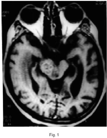
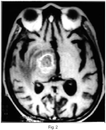
with 90% lymphocytes and 10% polymorphs. CSF proteins were 210 mg% and glucose 64%. MRI revealed
multiple conglomerate lesions in the midbrain (Fig.
1) and basal ganglia (Fig. 2), which were enhancing on
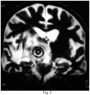
contrast. The image of interest is a noncontrast coronal MRI section, which showed a very peculiar target like
lesions with central dot and multiple hypo and hyperintense peripheral rings (Fig.
3). A diagnosis of tubercular
meningitis with multiple tuberculomas was made. The patient was started on antitubercular therapy.
Copyright 2002 - Neurology India.
Also available online at http://www.neurologyindia.com
The following images related to this document are available:
Photo images
[ni02110f1.jpg]
[ni02110f3.jpg]
[ni02110f2.jpg]
|
