 |
| About Bioline | All Journals | Testimonials | Membership | News |
|
||||||
|
||||||
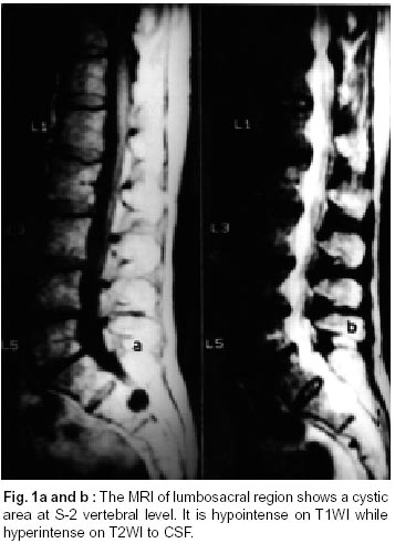
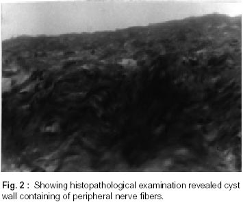
Neurology India, Vol. 50, No. 4, Dec, 2002, pp. 514-515 Short Report Sacral Perineural Cyst Presenting as Chronic Perineal Pain : A Case Report S.K. Jain, S. Chopra, H. Bagaria, P.P.S. Mathur Correspondence to : Dr. S.K. Jain, 436, Opposite Gudhas House, Haldiyon Ka Rasta, Johri Bazar, Jaipur - 302 003, Rajasthan, India. E-mail : shashi_neelu@yahoo.com Code Number: ni02138 Summary We present an interesting case of sacral perineural cyst which caused chronic perineal pain. Perineural cyst is relatively rare, especially the sacral region. Chronic perineural pain is an often encountered problem that is difficult to evaluate and sacral perineural cyst may be the etiology of chronic perineal pain in many instances. Key words : Perineal pain, Sacral Perineural cyst. Introduction Chronic perineal pain in medical practice is difficult to evaluate. Medical investigations including gynecological, urological and anorectal observations, fail to indicate a clear etiology in many of these cases and the perineal pain is often considered essential or of psychosomatic origin.1 Perineural cyst is relatively rare.2 The cyst within the sacral spinal canal, may be a sacral perineural cyst, sacral extradural cyst, occult intrasacral meningocele or anterior sacral meningocele.3 Sacral perineural cyst, was first described as an incidental autopsy finding by Tarlov in 1938.4 We are presenting an interesting case of sacral perineural cyst presenting as chronic perineal pain. Case Report A 55 year old female was admitted in the department of neurosurgery SMS Hospital, Jaipur with six months history of chronic perineal pain and intermittent claudication. There was no history of trauma. The pain, was most prominent in the coccygeal area and radiated to the pubic and vaginal regions. The neurological examination was normal including the evaluation of perineal region for touch, pain and temperature. Analgesic drugs and bed rest were of little benefit. Plain X-rays of lumbosacral region were normal. The MRI of lumbosacral region demonstrated a cystic area in the spinal canal at S-2 vertebral level with posterior scalloping of S-2 vertebra. It appeared hypointense on T1WI while hyperintense on T2WI (Fig. 1a and b). There was an incidental finding of primary spinal canal stenosis from L2-5 vertebra. LI to S3 wide decompressive laminectomy was performed and a cystic lesion on the right second sacral nerve root filling the whole sacral canal was found. The thecal sac, which ended proximally, was not involved or displaced by the lesion, which clearly compressed the other sacral nerve roots. The cyst wall was opened and removed after dissection of sacral nerve roots. Finally, the dural sheet was reconstructed. Histopathological examination revealed cyst wall containing peripheral nerve fibers (Fig. 2). Patient improved post operatively, and at the time of discharge had no perineural pain. Discussion Sacral nerve root cysts are commonly found during the third and fourth decades.5 Although most of patients are asymptomatic, some patients may have low back pain, sacrococcygeal pain, sensory and motor disturbances in lower extremities and urinary dysfunction with symptoms similar to lumbar disc prolapse3,6 The present case presented with chronic perineal pain with intermittent claudication. Much confusion exists in literature regarding the nomenclature for the various kinds of cysts of meninges. The origin of cysts has been the subject of debate. Spinal perineural cysts are considered to be congenital and are the diverticula of spinal meningeal sac, nerve root sheath and arachnoid.7 Considerable initial work has been done on this entity by Tarlov, who described extradural sacral cysts, often multiple and multilocular, usually found at the junction of posterior nerve root with the ganglion.8,9 He coined the term perineural cyst as he felt that these cysts emerged from the space between the endoneurium and perineurium as a result of ischemic degeneration, trauma or repeated hemorrhages beneath the perineurium of the root. Tarlov felt that these cyst had no communication with the subarachnoid space,8 which was also supported by Rexed and Wennstrom.10 Nabors et at11 showed that these cysts communicated with spinal subarachnoid space. Various mechanisms have been postulated to explain the progression in the size of the cyst such as hydrostatic pressure, osmotic pressure and secretory activity of cyst wall.12 Nabors et al11 introduced a classification system for sacral cysts. The spinal meningeal cyst can be classified in three major groups i.e. Type I - extradural cysts without spinal nerve roots, Type 2 - extradural cysts with spinal nerve roots and Type 3 - intradural cysts. MRI scan demonstrates sacral perineural cyst more frequently than CT scan with myelography. Further more, MRI scanning gives important information about the exact localization of the cyst. The sacral perineural cyst appears as hypointense to isointense signal intensity on T1WI and high signal intensity on T2WI,2 as in the present case. Microscopically the cyst wall consists of peripheral nerve fibers or ganglionic cells covered with meningeal epithelium,3 as in the present case. Bourgeois et al6 advocated two clinical types of radicular suffering from therapeutical point of view. Perineural cyst causing radicular extrinsic compression can be treated by surgery. The cystic nerve root can present an intrinsic suffering because of intradural dilaceration. Surgery must be avoided specially when many roots are involved because it may worsen the pleuriradicular suffering. Araki et al2 and Kopezynski et al13 reported relief of clinical symptoms after surgery as in the present case. References
Copyright 2002 - Neurology India. Also available online at http://www.neurologyindia.com The following images related to this document are available:Photo images[ni02138f1.jpg] [ni02138f2.jpg] |
| |||||||||
