 |
| About Bioline | All Journals | Testimonials | Membership | News |
|
||||||
|
||||||
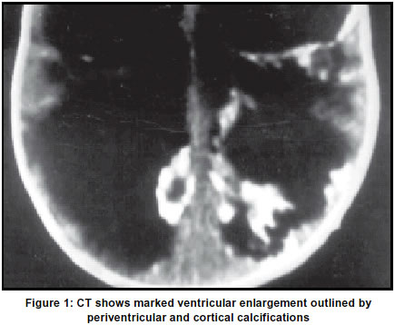
Neurology India, Vol. 51, No. 1, Jan-Mar, 2003, pp. 125 Letter to Editor Congenital toxoplasmosis infection M. B. Popli, V. Popli* Department of Radiological Imaging, Institute of Nuclear Medicine and Allied Sciences, New Delhi, India and *Department of Pediatrics, Maulana Azad Medical College and Associated Hospitals, New Delhi, India. Code Number: ni03042 Sir, A six-month-old child was brought to the hospital with complaints of delayed milestones and a large head. The child was born prematurely during the eighth month of gestation with a birth weight 2000 gm. Clinically, the patient was diagnosed as a case of hydrocephalus and a CT scan was performed (Figure 1). Choroidoretinitis was diagnosed on fundal examination. IgG was positive for toxoplasmosis. Toxoplasmosis is the disease caused by infection with the obligate intracellular parasite Toxoplasma gondii. This protozoon infection has been known for many years to infect animals and birds. Since 1942, it has also been known to infect humans. The cat is the definitive host and excretes the oocytes in its faeces. Infection in man occurs after ingestion of oocytes, or of tissue cysts in infected meat. In the persons whose immune system is intact, toxoplasmosis is usually asymptomatic and self-limited. This condition can go unrecognized in 80-90% of the adults and children with acquired infection. Unfortunately, a pregnant woman when infected can infect the fetus in utero.1 Congenital toxoplasmosis is an infection resulting from the transplacental passage of the parasites from an infected mother to the fetus. The infants born are usually asymptomatic at birth but later manifest a wide range of signs and symptoms including choroidoretinitis, strabismus, epilepsy and psychomotor retardation. The disease may have severe teratogenic effects on the fetal nervous system associated with intracranial calcification. There is marked brain necrosis and ventricles may be enormously dilated because of cerebral atrophy. Multiple calcified flecks are visible on CT scan. Toxoplasmic granulomata are widespread throughout the brain and often show marked calcification. Intracranial calcifications are scattered throughout the brain. They are usually clustered in the region of the choroid plexus and subependymal region or produce cast-like appearence of the ventricles. Calcification may also occur within the cortex as well as subcortical white matter. No calcifications are seen in the posterior fossa.2,3 32 cases of intracranial calcification secondary to toxoplasmosis were reported in 1968.4 Calcifications alone however, are not specific for toxoplasmosis as similar findings are seen with Cytomegalo virus and rubella (other TORCH infections). But typical ocular lesions with positive IgG titres establish the diagnosis of toxoplasmosis. M. B. Popli, V. Popli* Department of Radiological Imaging, Institute of Nuclear Medicine and Allied Sciences, New Delhi, India and *Department of Pediatrics, Maulana Azad Medical College and Associated Hospitals, New Delhi, India. References
Copyright 2003 - Neurology India. Also available online at http://www.neurologyindia.com The following images related to this document are available:Photo images[ni03042f1.jpg] |
| |||||||||
