 |
| About Bioline | All Journals | Testimonials | Membership | News |
|
||||||
|
||||||
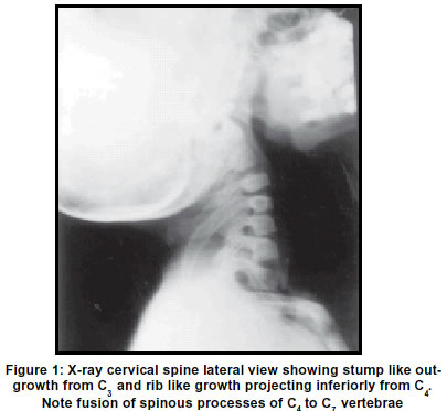
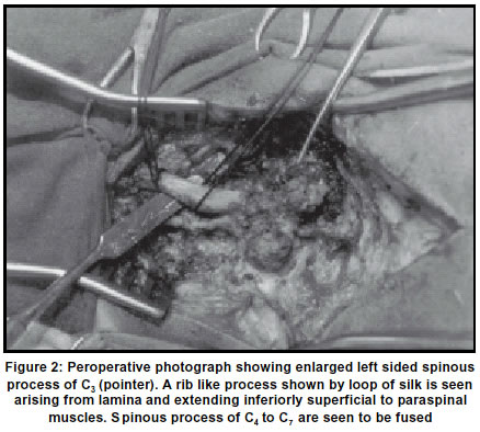
Neurology India, Vol. 51, No. 1, Jan-Mar, 2003, pp. 130-131 Letter to Editor Congenital exostoses of the cervical vertebrae N. Chitkara, N. K. Sharma, U. Dhall,* N. Bakshi, H. Kamal Department of Neurosurgery and *Anatomy, Pt. B.D.S. PGIMS, Rohtak, India. Accepted on 10.07.2001. Code Number: ni03047 Sir, Any osseous disease may involve the spinous processes. Since the tips of the spinal processes are palpable, their examination is sometimes of great diagnostic value. This applies not only to percussion pain, but also to other disorders of bone tissue which can be demonstrated by pressure and percussion. The superficial position of the tips readily permits a biopsy and has significance in the localization of vertebral levels. Congenital anomalies of the spinous processes might cause a problem in identification on the roentgenogram and preoperative localization. A 5-year-old female patient presented with two bony swellings at the back of the neck. The problem was only cosmetic to her. On examination, there was no neurological deficit. There was a bony stump palpable in the region of the upper cervical spine to the left of the spinous process. The second "rib"-like swelling was present beneath this, approximately 7 cm in length and running inferiorly to the left of the spinous processes of the lower cervical spine. X-ray cervical spine (Figure 1) confirmed these findings with no additional bony abnormality. MRI of the cervical spine showed normal spinal cord. Peroperatively, (Figure 2) "stump"-like bony growth was seen to be an enlarged left-sided spinous process of C3 vertebra. The inferior "rib"-like outgrowth had its origin from the lamina of C4 vertebra and was extending inferiorly up to D2 level. C4-C7 spinous processes were fused with normal muscle attachment on either side and the bony outgrowth was superficial to the muscle attachment. Both these outgrowths were excised from their base and on histopathological examination these had normal osseous structure. During 3-6 weeks of gestation, cellular differentiation, migration and segmentation of the axial skeleton takes place to form the mesenchymal analage of the vertebral column.1 Chondrification centers in this analage are established within 7 weeks of gestation2 and ossification centers appear from the second embryonic month onwards. The spinous process does not have its own ossification center (except for its tip), and is formed during the first year of life by fusion of the endochondral growing osseous extensions from both vertebral arches. The tip of the spinous process develops from a secondary ossification center at puberty. Hypoplasia of vertebral arches leading to spina bifida is well known. Hypoplasia of one arch and overgrowth of the other arch resulting in deviated spine is also known.3 Abnormal extension of chondrification and ossification of one vertebral arch with normal development of the other arch can only explain the present anomaly. Overgrowth of secondary ossification center for spinous process is ruled out, keeping in view the age of the patient. N. Chitkara, N. K. Sharma, U. Dhall,* N. Bakshi, H. Kamal Department of Neurosurgery and *Anatomy, Pt. B.D.S. PGIMS, Rohtak, India. References
Copyright 2003 - Neurology India. Also available online at http://www.neurologyindia.com The following images related to this document are available:Photo images[ni03047f1.jpg] [ni03047f2.jpg] |
| |||||||||
