 |
| About Bioline | All Journals | Testimonials | Membership | News |
|
||||||
|
||||||
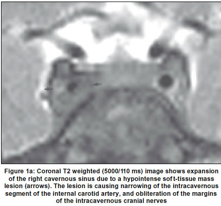
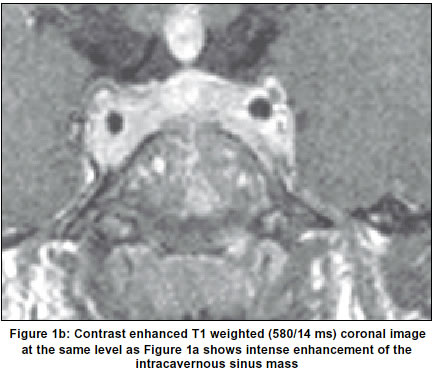
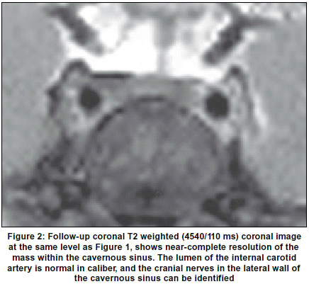
Neurology India, Vol. 51, No. 1, Jan-Mar, 2003, pp. 137 Neuroimage Tolosa-Hunt syndrome: MRI before and after treatment R. Koul, R. Jain* Department of Child Health (Neurology), Department of *Radiology, Sultan Qaboos University Hospital, Oman. Accepted on 24.05.2002. Code Number: ni03055 Contrast enhanced MRI of the brain revealed expansion of the right cavernous sinus due to an enhancing soft-tissue mass, engulfing and narrowing the intracavernous segment of the right internal carotid artery (Figures 1a & 1b). The child was started on oral Prednisolone 1 mg/kg/day. His headache resolved within 72 hours. The 6th nerve paralysis took 3 weeks to recover completely. Follow-up MRI after 6 weeks showed complete resolution of the abnormality (Figure 2). ESR at followup was 11 mm/ lst hour. Tolosa-Hunt syndrome1 is rare in children and is usually misdiagnosed as migraine which may not be so in adults.2,3 The condition is thought to be a non-specific inflammatory pseudotumor of the cavernous sinus. The most radiological differential diagnosis is from a cavernous sinus meningioma. The Tolosa-Hunt syndrome should be considered in severe and continuous headache with cranial nerve involvement (3rd, 5th and 6th) in children. MRI is the most important investigation and is diagnostic of the condition. References
Copyright 2003 - Neurology India. Also available online at http://www.neurologyindia.com The following images related to this document are available:Photo images[ni03055f1b.jpg] [ni03055f1a.jpg] [ni03055f2.jpg] |
| |||||||||
