 |
| About Bioline | All Journals | Testimonials | Membership | News |
|
||||||
|
||||||
Neurology India, Vol. 51, No. 2, April-June, 2003, pp. 241-243 Prevalence of photosensitivity - An Indian experience A. K. Roy, L. Pinheiro, S. V. Rajesh Department of Neurology, Command Hospital Airforce Hospital & St. John's Medical College Hospital, Bangalore. A. K Roy
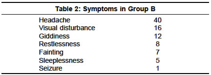
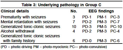
Accepted on 17.05.2001. Code Number: ni03073 ABSTRACT One thousand nine hundred and forty newly recruited entrants for training as pilots (Group A) underwent photic stimulation during EEG recording during the entrance examination to the flying stream. One hundred and sixty individuals (Group B) working on radars for prolonged periods were interviewed for eliciting complaints referable to photosensitivity and were subjected to EEG. EEGs in respect to 1000 cases (Group C) of known epilepsy were examined for the incidence of a photosensitive response. The study has revealed that 14 cases (0.72%) in group A had an abnormal response to photic stimulation out of which one case developed seizure during EEG recording. One case (0.62%) in group B and 30 cases (3%) in group C were detected to have photosensitivity. The maximum response was seen at 20 Hz stimulation. The prevalence of photosensitivity and its manifestations in these groups indicates that this condition is not uncommon. Key Words: Photosensitivity, Epilepsy, Prevalence. INTRODUCTION Epilepsy is said to affect 20-40 million people worldwide.1 About 5 % of patients with epilepsy show generalized paroxysmal epileptiform discharges in response to intermittent photic stimulation.2 Photosensitivity epilepsy has been described in 8% epileptic patients.3 In a prospective nationwide study in Britain, the annual incidence of the cases of epilepsy with a photo-paroxysmal response on their first EEG was conservatively estimated to be 1.1 per 100,000, representing approximately 2 % of all new cases of epilepsy. When restricted to the age range of 7-19 years, the annual incidence rose to 5.7 per 100,000, approximately 10 % of all new cases of epilepsy in this age range.4 There is a scarcity of Indian studies on the incidence or prevalence of photosensitivity epilepsy. Of 682 air force applicants studied by Buchtel and Lennox,5 5.1% showed a photo-paroxysmal response to intermittent photic stimulation with their eyes closed, whereas 2.6% had epileptiform discharges in their resting EEGs. The aim of our study was to determine the prevalence of photosensitivity (abnormal response to flickering light) in individuals with no previous exposure to flickering light, radar workers and in those with epilepsy. MATERIAL AND METHODS The prevalence of sensitivity to flickering light (photosensitivity) was studied in individuals by dividing the study population into three groups. Group A: (n=1940): Newly recruited entrants for training as pilots. Photic stimulation during EEG was done as part of basic medical examination. The number showing a photosensitive response was noted. Group B: (n=160): Individuals working on radars for prolonged periods were studied in a similar manner. Group C: (n=1000): Established cases of epilepsy were studied by intermittent photic stimulation, and their response in the form of photosensitivity was noted. Using the standardized method of IPS (intermittent photic stimulation), the photic stimulator was placed symmetrically in front of the patient's eyes, 30 cm from the nasion. The patient was seated and asked to look at the centre of the lamp. Standard room lighting was used as background illumination. The patient was asked to open the eyes; after 10 seconds, asked to close the eyes and IPS began at that moment at a series of standardized constant frequencies. The stimulus train lasted 10 seconds. During a 4-second pause, the eyes remained closed, and a further train of IPS was given for 4 seconds. The eyes were then opened for a 4-second period, with IPS (eyes-open) again given for a 4-second period. If a photo-paroxysmal response (PPR) occurred, there was a pause of 20 seconds, or at least four times the length of the discharge before the procedure was repeated at the next flash frequency. The test was started with 3 Hz stimulation, successively increased to 6,9,12 and 15 Hz. The upper limit was kept at 20 Hz and the flash frequency was decreased in a stepwise manner. The EEG responses found were: Regular spike and wave, or polyspike and wave activity with or without generalization. A "photo-myoclonic (PM) response" consisting of rhythmic action potentials from the orbital and other facial muscles synchronous with the photic stimuli and occurring only or predominantly in the frontal areas. RESULTS There were 1940 candidates who were newly recruited entrants for training as pilots (Group - A) (1930 males and 10 females). Their age ranged from 18-21 years. None of them had previous history of seizures or photosensitivity. Fourteen individuals (0.72%) had photo-convulsive (PC) response and of these, one person developed seizures during EEG recording. Photic-driving (PD) response was seen in 6 individuals and 2 individuals had photo-myoclonic (PM) response. In some patients, more than one type of abnormal response was seen. Of the 160 radar workers (Group B), there were 158 males and 2 females. Their age ranged from 20-50 years. They were grouped based on the duration of exposure and were interviewed for any neurological symptoms. It was noted that symptoms were more prevalent when the duration of exposure was prolonged (Table 1). Headache was the commonest symptom seen in 40 individuals. Other symptoms are summarized in Table 2. None of these individuals had previous history of seizures or photosensitivity. One person (0.62%) had a photo-convulsive response to IPS. He had been exposed to 6 hours of radar viewing for less than 1 year. One thousand known epileptics were subjected to IPS (723 males and 277 females, age range 8-20 years). Thirty cases (3%) had photosensitivity and maximum response was seen with stimulation at 20 Hz. The types of seizures and the EEG findings are summarized in Table 3. A photo-convulsive response was more commonly seen in those with idiopathic generalized tonic-clonic epilepsy (10 patients) and in those with mental retardation (7 patients). DISCUSSION Reflex epilepsies are defined as those seizures hat are reliably provoked by naturally occurring or artificial stimulation of a certain receptor or group of receptors.6 Photosensitive epilepsy is considered the main representative of the reflex epilepsies. Photosensitivity in humans is considered to be idiopathic and genetically transmitted.7 Of the typical photosensitive patients, 75% have tonic-clonic, myoclonic or absence seizures, whereas 25 % have a history of partial seizures. They typically have subtle eyelid myoclonic movements, jerks of the arms (mostly symmetric) and massive jerks of the whole body. These signs occur with or without alteration of consciousness. Visual aura is rare in these patients who, however, experience feelings of dizziness, light-headedness or warmth starting in the stomach and may find photic stimulation disagreeable.8 The most effective frequency of stimulation by IPS for evoking paroxysmal discharges is usually about 15-20 flashes/sec, but the range may be from 5-80 flashes/second.9 Screening methods using photic stimulation have been standardized.10 Bickford made a distinction between a photo-myoclonic response and a photo-convulsive response.11 Photo-myoclonus was a jerking response to light flashes in the facial muscles that could be detected in many normal subjects. In contrast, a clear generalized (poly) spike and wave discharge evoked by IPS (Intermittent Photic Stimulation) was strongly related to a history of epilepsy, especially when associated with myoclonic jerks. In a German epileptic population of 1000, 10% appeared to be photosensitive, using the criteria suggested by Bickford.12 Our study has revealed that 0.72% in the `normal' population had an abnormal response to photic stimulation, out of which one case developed seizure during EEG recording. The commonest finding in this group was photo-convulsive response. 0.62% of those with prolonged exposure to radars had photosensitivity. Many others had symptoms, the most common of which were headache and giddiness. 3% of known epileptics were detected to have photosensitivity. A photo-convulsive response was more commonly seen in those with idiopathic generalized tonic-clonic epilepsy and in those with mental retardation. In all 3 groups, cases with manifestations of photosensitivity were younger individuals. This is consistent with findings in previous studies.13 The maximum response to IPS was seen at 20 Hz. In conclusion, the prevalence of photosensitivity and its manifestations in these groups indicates that this condition is not that uncommon. Sensitivity to IPS (flickering) is an important cause of disability in individuals whose work environment exposes them to it, as it may precipitate seizures. In addition to the estimation of the prevalence of photosensitivity, IPS also helps to predict the relapse rate after partial or total withdrawal of the drug used in treatment (most commonly valproate). Eliciting symptoms of photosensitivity in epileptic patients is important, as it requires a different approach in the management. REFERENCES 1. Delgado-Escueta AV, Ward AA Jr, Woodbury DM, Porter RJ. New wave of research in the epilepsies. Adv Neurol 1986;44:3-55.
Copyright 2003 - Neurology India. Also available online at http://www.neurologyindia.com The following images related to this document are available:Photo images[ni03073t3.jpg] [ni03073t2.jpg] [ni03073t1.jpg] |
| |||||||||
