 |
| About Bioline | All Journals | Testimonials | Membership | News |
|
||||||
|
||||||
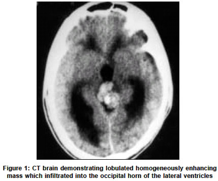
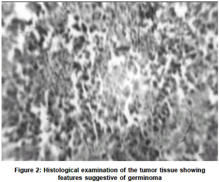
Neurology India, Vol. 51, No. 2, April-June, 2003, pp. 286 Letter to Editor Germinoma of the pineal gland S. C. Mohanty, A. K. Das, B. Tripathy, G. Mohanty, S. Bhagat, S. Mishra Departments of Neurosurgery, Neurology, Radiology and Pathology, V. S. S. Medical College, Burla-768017, Orissa. Accepted on 17.09.2002. Code Number: ni03095 Sir, A 17-year-old boy presented with generalized headache, vomiting, ataxic gait and worsening of vision of 3 months duration. The onset was insidious and the course was progressive. Fifteen days prior to hospitalization, he developed bilateral ptosis, upward gaze palsy and bilateral gross papilloedema. Cerebellar and frontal lobe signs were absent. Plain radiography of the skull showed fluffy calcification in the pineal region. Plain CT brain demonstrated an isodense mass in the posterior third ventricle with calcification and obstructive hydrocephalus involving the third and lateral ventricles. The tumor enhanced homogeneously on contrast administration. It was lobulated and infiltrated into the occipital horn of the lateral ventricles (Figure 1). A right-sided ventriculoperitoneal shunt was performed. Following shunt insertion, the patient improved symptomatically. On the 10th postoperative day, the tumor was approached through the supracerebellar infratentorial route [Krause's approach] in park bench position. The precentral and superior vermian veins were coagulated and the tumor was visualized. The tumor was grayish in colour, soft and moderately vascular. Near total excision of the tumor was done. The postoperative period was uneventful. Histological examination of the tumor tissue confirmed the diagnosis of germinoma (Figure 2). He was subjected to a course of radiotherapy, 3600cGY to the ventricular system and 1900cGY to the tumor bed. At follow-up after 2 months, the patient had recovered from Parinaud's syndrome. At 2 years following surgery, the patient is asymptomatic and leading a normal life. CT brain showed no evidence of tumor. There are few studies documenting the long-term survival of patients with pineal tumors. Germinomas represent a rare category of malignant tumors for which treatment can be curative. Long-term survival with surgery and radiation is in the order of 75 to 80%. REFERENCES
1. Bruce JN. Management of pineal region tumours. Neurosurg Q 1993;3:103-9. Copyright 2003 - Neurology India. Also available online at http://www.neurologyindia.com The following images related to this document are available:Photo images[ni03095f1.jpg] [ni03095f2.jpg] |
| |||||||||
