 |
| About Bioline | All Journals | Testimonials | Membership | News |
|
||||||
|
||||||
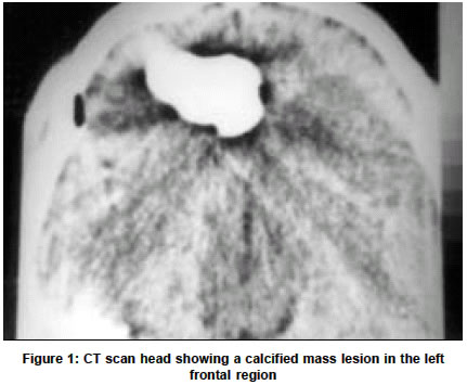
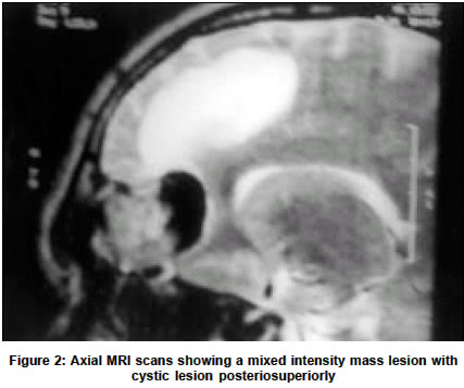
Neurology India, Vol. 51, No. 2, April-June, 2003, pp. 287 Letter to Editor Osteoma mimicking a partly calcified meningioma N. Chitkara, N. K Sharma, N. Goyal Accepted on 27.06.2002. Code Number: ni03096 Sir, Osteomas, as the name implies, consist of dense compact bone. They arise in bone formed by intramembranous ossification and are slowly growing neoplasms composed of mature lamellar bone.1 However, these may occasionally have an admixture of osseous and fibrous elements.2 A 25-year-old male presented with generalized seizures for 2 months. Neurological and systemic examination was normal. CT scan (Figure 1) showed a calcified lesion in the left frontal region. In addition there was a pneumocephalus in the temporal region. MRI scan (Figure 2) showed an extra axial mass lesion in the floor of the left anterior fossa which was of mixed intensity anteriorly and low intensity posteriorly. The ethmoid sinus of that side was seen to be compressed. In addition, there was a cystic lesion in relation to the tumor extending posteriorly up to the frontal horn of the lateral ventricle. Frontal craniotomy and excision of the tumor was done which on histopathology was confirmed to be an osteoma. Osteomas are the most common primary benign bone tumors found in the craniofacial skeleton.1 These tumors in the skull commonly arise within the paranasal sinuses and are most frequently located in the frontal sinus (40-80%).2 They usually expand within and conform to the shape of the sinus. Intracranial extension is rare because osteomas of the vault tend to arise from the outer table and grow outward. Pneumocephalus has been reported as a complication of frontal and ethmoidal osteomas after they have eroded the floor of the anterior cranial fossa.2 This feature serves to differentiate a mixed density osteoma from other intracranial mass lesions like meningioma as in this case. References 1. Hamilton HB, Voorhies RM. Tumors of the skull. In: Wilkins RH, Rengachary SS, eds. Neurosurgery. New York: McGraw-Hill; 1996. pp. 1503-28. Copyright 2003 - Neurology India. Also available online at http://www.neurologyindia.com The following images related to this document are available:Photo images[ni03096f1.jpg] [ni03096f2.jpg] |
| |||||||||
