 |
| About Bioline | All Journals | Testimonials | Membership | News |
|
||||||
|
||||||
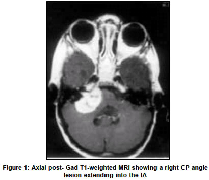
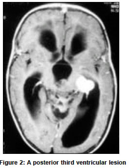
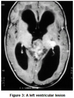
Neurology India, Vol. 51, No. 2, April-June, 2003, pp. 297-298 Neuroimage Multifocal intracranial rhabdoid tumor A. Suri, V. P. Singh, S. S. Kale, V. S. Mehta, S. Gaikwad* Departments of Neurosurgery and *Neuroradiology, Neurosciences Center, All India Institute of Medical Sciences, Ansari Nagar, New Delhi-110029, India. A. Suri Accepted on 16.01.2001. Code Number: ni03106 An 18-months-old boy presented with progressive inability to close the right eye, deviation of angle of the mouth and difficulty in standing and sitting without support for a period of 2 months. Examination revealed an irritable and uncooperative child with a head circumference of 51 cm. The anterior fontanel was full but not tense. Examination of the fundus revealed bilateral mild papilloedema. There was right-sided lower motor neuron facial palsy. Mild truncal ataxia was present with a tendency to fall on the right side. MRI revealed a right cerebello-pontine (CP) angle lesion extending into the internal auditory meatus (IAM) (T1 - hypo, T2 - hyper, homogeneously enhancing), a posterior third ventricular and superior vermian lesion (T1 - hypo, T2- iso, heterogeneously enhancing), and a left ventricular lesion (T1 -hypo, T2 - hypo, homogeneously enhancing) with moderate ventriculomegaly The child underwent left ventriculoperitoneal shunt following which he was operated for the right CP angle (retromastoid suboccipital approach) and posterior third ventricular tumor (suboccipital supracerebellar approach) in sitting position in a single operation. The child did well for 36 hours postoperatively after which he had left lateral ventricular bleed to which he succumbed. Histology and electron microscopy revealed similar pathology at both locations rhabdoid tumor, i.e. atypical teratoid tumor. DISCUSSION Rhabdoid tumor (atypical teratoid tumor) is a unique combination of neuroepithelial, peripheral epithelial and mesenchymal elements and histologically, is often confused with meduloblastoma / primitive neuroectodermal tumor (PNET). Age at onset is less than 3 years (median age 10.5 months) with male preponderance. They are very uncommon intracranial tumors. Rare features that highlight this case are multiple tumors in the supratentorial as well as infratentorial compartments with different radiological characteristics and presentation at the CP angle with extension into the IAM. They are highly aggressive tumors with a median survival time of 6 months despite intensive therapy. The recognition of this entity is very essential for aggressive management and prognostication of the patient, which is drastically different from that for PNET. Copyright 2003 - Neurology India. Also available online at http://www.neurologyindia.com The following images related to this document are available:Photo images[ni03106f3.jpg] [ni03106f1.jpg] [ni03106f2.jpg] |
| |||||||||
