 |
| About Bioline | All Journals | Testimonials | Membership | News |
|
||||||
|
||||||
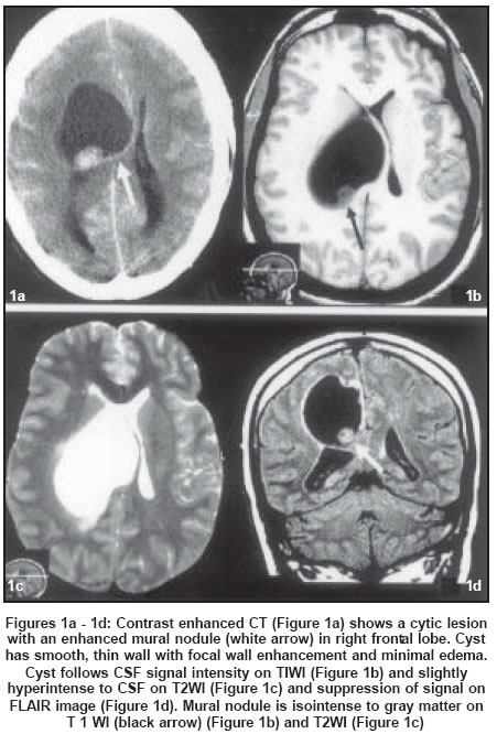
Neurology India, Vol. 52, No. 1, January-March, 2004, pp. 136 Neuroimage Cyst with a mural nodule: Unusual case of brain metastasis Garg A, Suri A, Gupta V Department of Neurosurgery, AIIMS, New Delhi Code Number: ni04050 A 34-year-old woman presented with history of headaches, vomiting and progressive left hemiparesis for the last 3 months. CECT showed right frontal juxtaventricular, cystic lesion with an enhancing mural nodule and focal wall [Figure:1a]. On MRI, the cyst was hypointense on T1WI [Figure:1b] and hyperintense onT2WI [Figure:1c and suppressed on FLAIR sequence [Figure:1d]. Per-operatively, a cyst with yellowish clear fluid and a small grayish, solid part were removed. The cyst walls were not necrotic. HPE revealed a malignant tumor composed of cells with focal areas of glandular differentiation with marked pleomorphism. The tumor cells were immunoreactive for cytokeratin and mucin. The wall of the cyst showed gliosis. A diagnosis of metastatic adenocarcinoma was made. No primary could be localized. The radiological differential considerations for a cystic tumor with an enhancing mural nodule include pilocytic astrocytoma, hemangioblastoma, pleomorphic xanthoastryocytoma, meningioma and ganglioglioma.[1],[2] The radiological finding of a cystic tumor with a mural nodule had not been described previously in brain metastases. The presence of minimal edema relative to the size of the lesion in our case was also unusual for a metastatic deposit. FLAIR sequences have been reported to be useful in distinguishing between cystic neoplastic and non-neoplastic lesions.[3] The cyst had suppression of signal intensity on FLAIR imaging, thereby suggesting non-mitotic pathology. This unusual appearance is possibly attributed to the lack of proteinaceous or myxoid material inside the cyst, which is rare in metastasis. Therefore, FLAIR images should be interpreted with caution. References
Copyright 2004 - Neurology India The following images related to this document are available:Photo images[ni04050f1.jpg] |
| |||||||||
