 |
| About Bioline | All Journals | Testimonials | Membership | News |
|
||||||
|
||||||
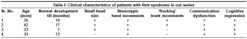
Neurology India, Vol. 52, No. 4, October-December, 2004, pp. 494-495 Case Report Recent experience with Rett syndrome at a tertiary care center Kumar Sudhir, Alexander M, Gnanamuthu C Department of Neurological Sciences, Christian Medical College, Vellore - 632004 Code Number: ni04165 ABSTRACT Rett syndrome (RS), a neurological developmental disorder, is one of the commonest causes of cognitive impairment in girls and women. These patients are often initially misdiagnosed as idiopathic mental retardation, cerebral palsy, or autism. Despite several reports from the West, there are very few reports from the Indian population. We present four female children with RS and emphasize the importance of early diagnosis.Key Words: Rett syndrome, Mental retardation, Clinical diagnosis. INTRODUCTION Rett syndrome (RS), one of the commonest causes of profound cognitive impairment in girls and women, was first recognized in the 1960s and the first report form India was in 1994.[1] Though RS has a prevalence of 1 per 10000 to 1 per 22000,[2] in India it is confined to very few case reports.[3],[4] Probably, this may be related to underdiagnosis of the condition or misdiagnosis of these children as cerebral palsy[1] or autism. The current report summarizes our recent experience with RS and discusses the clinical diagnosis. CASE REPORT Retrospective review of the case records of inpatients in the neurology wards during the period 2001-2002, identified four female children with RS. The mean age at presentation was 33 months (23-42 months). Diagnosis was based on the criteria proposed by Hagberg and Witt-Engerstrom.[6] The clinical characteristics are summarized in [Table - 1]. All children had uneventful antenatal and perinatal periods and a period of normal development, ranging from the first seven to 18 months. During the period of normal development, three children were able to walk without support. They also attained a vocabulary of 8-10 words. Family history was negative in all. Later in the course of illness, the complaints were an inability to use hands for purposeful movements, social withdrawal, problems with communication and regression of acquired motor and language skills. They had marked communication problems and the vocabulary regressed to one-two words in three children and four-six words in one. They had difficulty in comprehending spoken language but could respond to loud noise and bright light. Stereotypic hand movements were noted in all the children. The stereotypic movements included beating hands on the abdomen in one (Case 1), repeated closing and opening of hands in one (Case 2), slapping own face with hands and wringing movements in one (Case 3) and repeated inappropriate hand clapping in one (Case 4). Gait problems, apraxia, was noted in all the children. One child (Case 1) had mild swaying to either side, whereas 2 children (Cases 2 and 3) had broad-based unsteady gait with repeated falls. One child (Case 4) had an unusual way of turning to one side before getting up from the squatting position to avoid falling. Side-to-side ′rocking′ was seen in two children (Cases 1 and 2). All children had cognitive impairment and had no bowel and bladder control. Two patients had history of seizures. One child (Case 1) had myoclonic jerks whereas another child (Case 4) had generalized tonic-clonic seizures. Two patients had bruxism. One patient had hearing loss. Three children (Cases 1, 2, and 3) had microcephaly. Weight and height were below the 50th percentile in all the four children. None had any dysmorphic features. Muscle wasting and hypotonia was noted in one child and one child had spasticity. Brain CT was normal in three children, in Case 2 MRI showed frontal lobe atrophy and thinning of the anterior corpus callosum. EEG was abnormal in all the four children (multifocal epileptiform activity in Cases 1 and 3 and focal epileptiform discharges in Cases 2 and 4). CSF analysis including measles antibody titer was normal. Genetic analysis was not performed in our cases. DISCUSSION Rett syndrome is characterized by profound cognitive impairment, problems with communication, stereotypic hand movements, and pervasive growth failure that follow a normal period of development during the first six to 18 months of life.[2] Diagnosis of RS is based on clinical criteria,[5] as only 70-80% of patients with typical RS phenotype have mutations in the RS gene. Our patients fulfilled all the essential and exclusion criteria. Supportive criteria noted in our patients were bruxism (50% of patients) and growth retardation (100% of patients). Clinical features of RS are classified into four stages.[6] First stage is the stagnation period with onset between six and 18 months of age. It lasts for weeks to months and is characterized by developmental delay without regression. Second stage starts between one and four years of age and is characterized by regression of motor and language milestones. Stage Three or pseudostationary stage may last for several decades and is seen in girls with preserved ambulation. Communication may even improve but the motor functions worsen. Stage four is characterized by loss of ambulation. The transition from one stage to the next does not occur abruptly. All our cases were seen in the first two stages. Seizures have been reported in 30-94% of patients with RS. In a series of 53 females with RS, 94% had a history of epilepsy, focal epilepsy being twice as common as generalized.[7] The mean age of onset of epilepsy was significantly higher as compared to the onset of cognitive impairment (4 years versus 0.8 years). Two of our patients had epilepsy. Bruxism is one of the most common manifestations in RS.[8] The prevalence of sensorineural and conductive hearing loss was reported in 17% and 10% respectively.[9] Bruxism was seen in two and hearing loss in one of our patients. Rett syndrome is commonly misdiagnosed as cerebral palsy.[1] Clinical features distinctive for RS, however, include normal antenatal and perinatal periods, a normal period of development for the first 6-18 months of life and regression of acquired milestones. RS is also misdiagnosed as autism. Autistic children lack social skills and prefer objects to people, whereas RS girls prefer affection and company of people.[10] Though autistic features may be present initially in RS, they tend to decline with age. Neuroimaging studies in RS have shown generalized cerebral atrophy predominantly affecting the frontal lobes.[11] EEG abnormalities are reported in almost all cases and findings vary with the age of the patient and the clinical stages of RS.[2] EEG was abnormal in all our cases. Rett syndrome is a genetic disorder transmitted in an X-linked dominant fashion, 99% of the cases are sporadic, and familial recurrence is seen in less than 1%.[3] Mutations in the MECP2 gene, located in the region Xq28, have been identified in 70-80% of patients.[12] Genetic studies are not essential for diagnosis in the majority, children with mild or atypical symptoms may require molecular genetic studies. Angelman syndrome and RS may have phenotypic and EEG similarities. In such cases molecular genetic studies may differentiate the disorders. Treatment is symptomatic and supportive. Long-term management involves physical and occupational therapy, speech therapy, nutritional support, seizure control and orthopedic intervention. Patients need scoliosis surveillance and surgery is recommended when the curve passes 40 degrees, which is associated with good outcome.[13] Feeding problems may benefit with changing the food texture and may require alternate routes of feeding in some cases. In conclusion, RS is probably underdiagnosed in our country and should be suspected in appropriate clinical situations as the diagnosis can be made clinically. It should be differentiated from cerebral palsy and autism, as the therapeutic approach and outcomes differ. REFERENCES
Copyright 2004 - Neurology India The following images related to this document are available:Photo images[ni04165t1.jpg] |
| |||||||||
