 |
| About Bioline | All Journals | Testimonials | Membership | News |
|
||||||
|
||||||
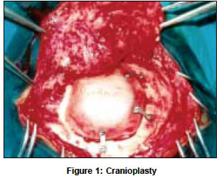
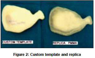
Neurology India, Vol. 52, No. 4, October-December, 2004, pp. 520 Letter To Editor Custom cranioplasty using rapid prototyping technology Parthiban JuttyKB, Abirami O, Murugan ArulM, Radhakrishnan R Departments of Neurosurgery, Kovai Medical Centre Hospital, PSG College of Technology, Peelamedu, Coimbatore - 641004 Code Number: ni04182 Sir, Cranioplasty for a large skull bone defect can be a challenging surgical problem. A variety of materials have been used for cranioplasty. Due to the advances in bioengineering, custom templates and prosthesis are now available for cranioplasty using Rapid Prototyping (RP) technology. Rapid prototyping technology was originally developed to rapidly build a prototype of a new product, especially in automobile industry. Now the same technology is used in the medical field in the production of anatomical models and templates, which facilitate surgeons to optimize preoperative surgical planning, interactive surgical simulation, while reducing operative time and complications. TIFAC CORE (Technology Information, Forecasting and Assessment Council - Center of Relevance and Excellence), PSG College of Technology, Coimbatore provided the first custom template for cranioplasty in our patient. Under the broad umbrella of technology vision-2020, TIFAC has set up a center of Relevance and Excellence in product design, optimization and collaborative product.[1] A frontotemporal craniectomy defect in a head injury victim was closed with a prosthesis made up of a biocompatible substance polymethyl metha acrylate [Figure - 1]. This prosthesis was a replica of the custom template produced by Rapid Prototyping Technology, using the data of 3D-CT scan images and 2 mm CT cuts submitted to TIFAC CORE [Figure - 2]. Rapid prototyping (RP) is a term that has been used to describe the production of solid models from 3D computer data by a group of relatively new technologies.[2] Using an additive approach to building shapes, RP systems join liquids, powder or sheet materials to form physical objects layer by layer. Rapid prototyping is now widely applied in the medical field. Some of the classical fields for medical applications of RP models are surgical planning and simulation, surgical rehearsal, training of student surgeons and radiologists, communication between medical staff and patients, and design of individual implant and prostheses. Some of the common additive Rapid prototyping technologies in medicine are Selective Laser Sintering (SLS), Fused Deposition Modeling (FDM), Stereolithography and recently, the most advanced Multi-jet Modeling.[3] Rapid prototyping technology has shown significant benefit in Maxillo facial reconstruction, cranio synostosis, skull and maxillo facial tumor surgery, skull plasties, orthodontic surgery, deformities of long bone joints and knee surgery, pelvic fractures, hip dysplasia, spinal trauma, congenital and degenerative spinal diseases, foot and hand malformations, and in models of soft tissue structures such as the cardiovascular system. The most exciting case in which RP technology was used in the recent past was in planning the successful separation of conjoined twins (Siamese twins) by using the RP model of the twins′ brain and their venous structure. REFERENCES
Copyright 2004 - Neurology India The following images related to this document are available:Photo images[ni04182f1.jpg] [ni04182f2.jpg] |
| |||||||||
