 |
| About Bioline | All Journals | Testimonials | Membership | News |
|
||||||
|
||||||
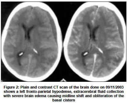
Neurology India, Vol. 52, No. 4, October-December, 2004, pp. 522-523 Letter To Editor Fulminant subdural empyema-an unusual complication of pyogenic meningitis Shenoy SN, Rao SN, Raja A Department of Neurosurgery, Kasturba Medical College and Hospital, Manipal - 576 119, Udupi, Karnataka Code Number: ni04185 Sir, An otherwise normal 56-year-old diabetic patient presented with a one-day history of multiple generalized tonic-clonic seizures followed by altered sensorium. There was no history of fever. There was no history of trauma or any focus of infection. There was no focal neurological deficit or signs of meningitis. Hematological investigation revealed leucocytosis with a total white blood cell count of 16,600/cu.mm and an ESR of 45 mm. Blood sugar was 351 mg%. Computed tomography (CT) scan of the brain revealed no abnormality [Figure - 1]. The lumbar CSF analysis revealed 1350 cells/mm with 96% neutrophils and 04% lymphocytes. Blood and CSF cultures did not reveal any growth. A diagnosis of pyogenic meningitis with diabetes was considered and the patient was placed on broad-spectrum antibiotics. Two days after admission to the hospital she developed recurrent attacks of seizures, lapsed into altered sensorium and developed a left pupillary dilatation. Repeat CT scan revealed a left fronto-parietal hypodense, extracerebral fluid collection with severe brain edema causing midline shift and obliteration of the basal cistern [Figure - 2]. An emergency left frontal burr hole was done and thick pus was evacuated. Gram′s stain revealed pus cells and gram-negative bacteria and the culture showed growth of Klebsiella species. The patient deteriorated rapidly and died. Subdural empyema complicating meningitis is relatively common in infants, but is rare in adults.[1],[3] Our experience in the present case suggests that subdural empyema should be suspected in patients with pyogenic meningitis who develop recurrent seizures, focal neurological deficit or deteriorate neurologically.[3],[4],[5] REFERENCES
Copyright 2004 - Neurology India The following images related to this document are available:Photo images[ni04185f2.jpg] [ni04185f1.jpg] |
| |||||||||
