 |
| About Bioline | All Journals | Testimonials | Membership | News |
|
||||||
|
||||||
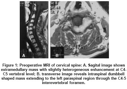
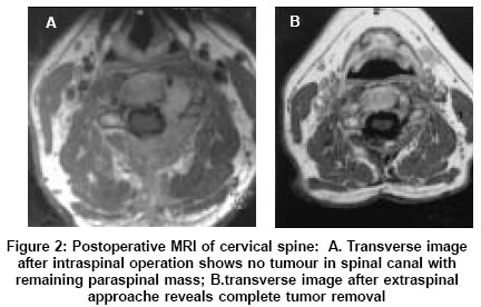
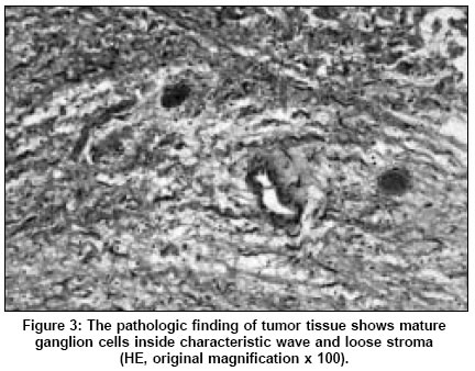
Neurology India, Vol. 53, No. 3, July-September, 2005, pp. 370-371 Letter To Editor Cervical dumbbell ganglioneuroma producing spinal cord compression Radulovi DaniloV, Branislav D, Skender-Gazibara MilicaK, Igor NikoliM Institute for Neurosurgery, Belgrade Date of Acceptance: 19-Apr-2005 Code Number: ni05134 Sir, A 39-year old man presented with complains of progressive weakness and numbness of all four limbs for six months. There was moderate spastic tetraparesis that was more marked on the left side, and hypoesthesia below the C5 dermatome. Magnetic resonance imaging (MRI) showed a large extramedullary dumbbell mass at the C4-C5 level. The tumor was hypointense on the T1 and hyperintense on T2 images. The spinal cord was severely compressed [Figure - 1] A and B. A two-staged operation was performed to resect the tumor. First, the patient was operated through a posterior cervical approach. Wide laminectomy of C4 and C5 was done. The mass was solid, well capsulated, elastic, moderately vascularised, purely extradural and ventrolaterally located to the spinal cord. The lesion originated from cervical nerve C5, which was resected with tumor [Figure - 2]A resection of intraforaminal mass was performed through the foramen which had been already enlarged by tumor growth. After four weeks the patient underwent second operation. The paraspinal extravertebral component of tumor was excised through the left lateral cervical approach [Figure - 2]B. The vertebral artery was dissected off the surface of encapsulated tumor. At a three-year follow-up the patient had regained the motor strength in all four limbs. There was no radiographic signs of recurrence. Histological examination of both tumor masses confirmed that the lesion was a ganglioneuroma [Figure - 3]. Discussion Kyoshima et al[1] surveyed the literature on the subject and identified a total of only five pathologically confirmed cases of cervical spine ganglioneuromas. One patient was an 18-month old child and rest of the patients were young adults. Von Recklinghausen′s disease was present in two patients. The symptoms spinal cord compression were present in all reported cases. Two patients had bilateral tumors. The origin of tumors was sensory root ganglion or cervical nerve. In all the reported cases, the tumor growth was in dumbbell pattern. Intraspinal extradural growth was observed in three patients, while intradural extension was seen in two patients. On MRI about 75% of ganglioneuromas are isointense and 25% are hypointense on T1 images. Most of them are hyperintense on T2 images. The non-homogeneous appearance corresponds to areas of cystic degeneration, hemorrhage or necrotic degeneration.[4],[5] Ganglioneuromas are well encapsulated tumors and can be completely excised. Even when they are intradural, the tumor could be removed without cord injury because they are not adherent to the spinal cord.[1] This and previously reported cases indicate that spinal ganglioneuromas could be completely removed and cured. References
Copyright 2005 - Neurology India The following images related to this document are available:Photo images[ni05134f3.jpg] [ni05134f1.jpg] [ni05134f2.jpg] |
| |||||||||
