 |
| About Bioline | All Journals | Testimonials | Membership | News |
|
||||||
|
||||||
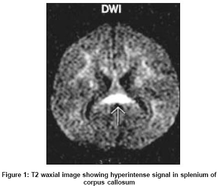
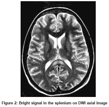
Neurology India, Vol. 53, No. 3, July-September, 2005, pp. 377-378 Neuroimage Hyperintense splenium in vitamin B12 deficiency Kori S Dept Of Radiodiagnosis K.E.M.Hospital, Pune 411011 Date of Acceptance: 24-Mar-2005 Code Number: ni05140 A 15-year-old female vegan was hospitalized for 6 days of fever, abdominal pain, and loose stools. The patient was found to have gross difficulty in walking, normal higher functions with MMSE of 28/30, absence of apraxia, dysarthria, and nystagmus; she had normal power in the limbs, normal deep jerks, and negative Babinski response. The touch, pain, joint position, and vibration sense were grossly normal; she showed severe finger to nose and gait ataxia. The Romberg sign was unequivocally and strongly positive and her gait ataxia would worsen on eye closure. The laboratory values were: hemoglobin: 5.7 g%; packed cell volume: 14%; mean corpuscular volume: 109 fl; red blood cell count: 1.34 x 10⁶sub/ml; white blood cell count: 1.9 x 10³sub/ml; platelets: 4.1 x 10³sub/ml; peripheral smear showed macrocytes and macro-ovalocytes; LDH: 918 U/l (N 90-200 U/l); serum vitamin B12: 78 pg/ml (N 200-950 pg/ml); serum folic acid: 0.81 ng/ml (N 3-17 ng/ml); serum homocysteine: 32 μl/l (N 2-7 μl/l). The results of the thyroid, liver, and renal function tests were normal. The work-up for collagen vascular disease, malabsorption and for fever was negative. The nerve conduction velocities were normal, save for absent F responses from the tibial and peroneal nerves on both sides. The lumbar puncture was deferred, as the platelet count was low. The T2 and diffusion weighted images (DWI) of brain magnetic resonance imaging showed hyperintensity in the splenium [Figure - 1] and [Figure - 2]. Subtle hyperintense signal was noted in bi-frontal and parietal white matter on T2W images. The magnetic resonance imaging of cervical spine showed posterior column hyperintensity extending from C2 to C5 vertebrae on T2W images, both in sagittal and axial planes. The acute presentation of sensory ataxia was likely to be due to aggravation of the nutritional deficiency of vitamin B12 by the febrile illness. The MRI was repeated 1 month after injectable and oral cyanocobalamine treatment, and it showed persistence of the hyperintense signal in the splenium; however, its intensity was reduced. Hyperintense signal from splenium of corpus callosum has been observed in hypoxic-ischemic encephalopathy, Marchiafava-Bignami syndrome,[1] high-altitude cerebral edema,[2] antiepileptic drug toxicity,[3] following epileptic attacks,[4] radiation therapy,[5] hemolytic uremic syndrome,[6] CNS lymphoma, astrocytoma, and demyelinating disease.[7] The hyperintense signal from the splenium of corpus callosum in vitamin B12 deficiency has not hitherto been described. At follow-up after 3 months, the patient was asymptomatic. References
Copyright 2005 - Neurology India The following images related to this document are available:Photo images[ni05140f2.jpg] [ni05140f1.jpg] |
| |||||||||
