 |
| About Bioline | All Journals | Testimonials | Membership | News |
|
||||||
|
||||||
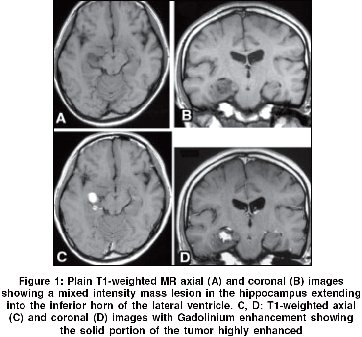
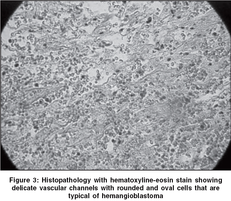
Neurology India, Vol. 54, No. 1, January-March, 2006, pp. 89-90 Case Report Hemangioblastoma of hippocampus without von Hippel-Lindau disease: Case report and review of literature Ohata K, Takami T, Tsuyuguchi N, Hara M, Haque M Department of Neurosurgery, Graduate School of Medicine, Osaka City University, Osaka Code Number: ni06025 Abstract A rare case of hemangioblastoma located in the region of hippocampus is reported. A 27-year-old female presented with a single episode of generalized convulsion. The vascular and cherry red color hemangioblastoma was resected by a temporo-zygomatic approach. There has been no recurrence of tumor at a follow-up of 11 yearsKeywords: Cerebrum, hemangioblastoma, hippocampus Supratentorial hemangioblastomas are rare tumors. From 1902 to 2005, around 118 cases (excluding our case) have been reported in the literature.[1],[2],[3],[4],[5],[6],[7],[8],[9],[10] These tumors originated in various supratentorial locations with or without associated von Hippel-Lindau disease. We could not locate any report of hemangioblastoma located in the region of hippocampus. The management of the case is reported. Case Report The 27-year-old right-handed female presented with a single episode of generalized convulsion. Fundoscopic examination, ultrasonography of the abdomen and the family history did not reveal any clinical feature of von Hippel Lindau disease. Magnetic resonance imaging (MRI) showed a mixed intensity solid mass lesion in the right hippocampal gyrus extending in to the temporal horn of the lateral ventricle. The lesion measured 18 x 12 x 15 mm in size. The lesion enhanced on Gadolinium administration [Figure - 1]. Conventional angiography showed that the tumor was highly vascularized and a branch of the anterior choroidal artery fed the tumor [Figure - 2]. A cherry red highly vascular tumor was totally removed through a temporo-zygomatic approach on 21st October, 1994. Histology confirmed that the lesion was a hemangioblastoma [Figure - 3]. The postoperative course was uneventful. At a follow-up after 11 years, the patient is asymptomatic and investigations have revealed no tumor recurrence or presence of any other mass in the central nervous system or in any other organ. Discussion Approximately 118 cases of supratentorial hemangioblastomas have been reported in the literature. The tumors have been identified in a variety of locations of the brain.[1],[2],[3],[4],[5],[6],[7],[8],[9],[10] In the analysis of 80 cases, the incidence of tumor in location depended on the volume of parenchyma of each location. The age ranged from 3- to 83-year old (average age 36.2 years) and there was a male predomination.[1] Association with von Hippel-Lindau disease is estimated to be 16% of all supratentorial hemangioblastomas,[1] which is almost similar to the coincidence (13.5-34.3%) of cerebellar hemangioblastomas and von Hippel-Lindau disease.[1] Although 13 cases of hemangioblastoma have been identified in the temporal lobe,[1],[2], [5],[6],[7],[8],[9],[10] hemangioblastoma limited to the hippocampus is not reported. To our knowledge, this is the first case report of hemangioblastoma of the hippocampus. References
Copyright 2006 - Neurology India The following images related to this document are available:Photo images[ni06025f3.jpg] [ni06025f1.jpg] [ni06025f2.jpg] |
| |||||||||
