 |
| About Bioline | All Journals | Testimonials | Membership | News |
|
||||||
|
||||||
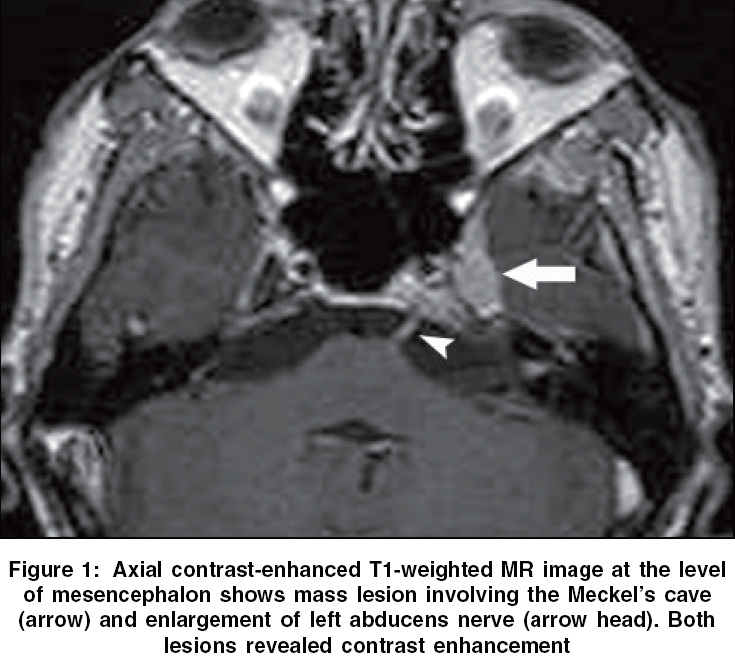
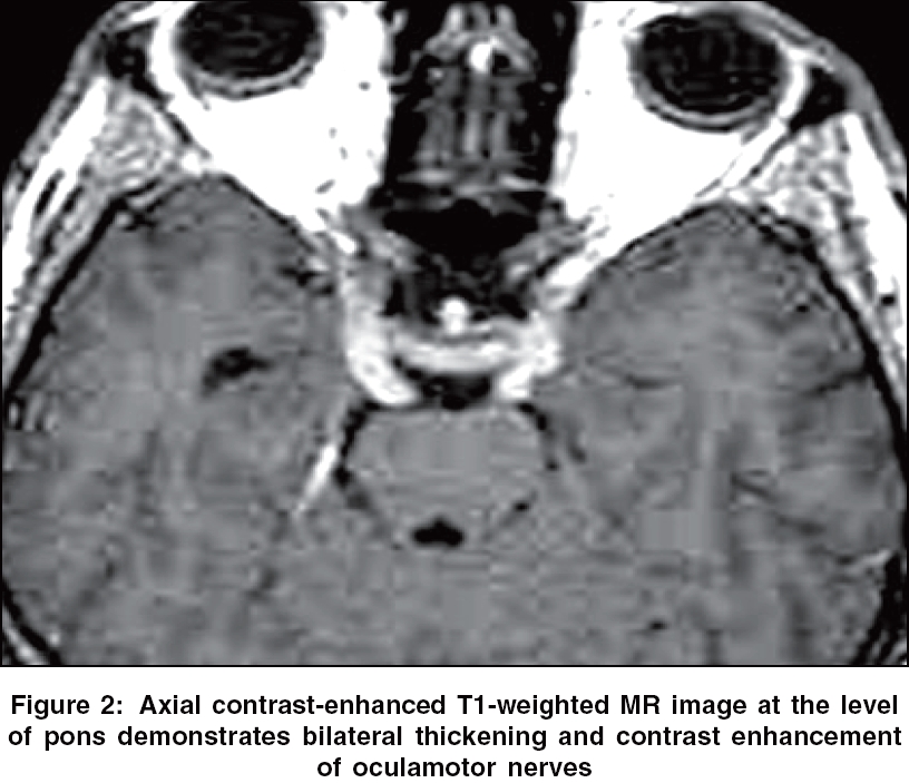
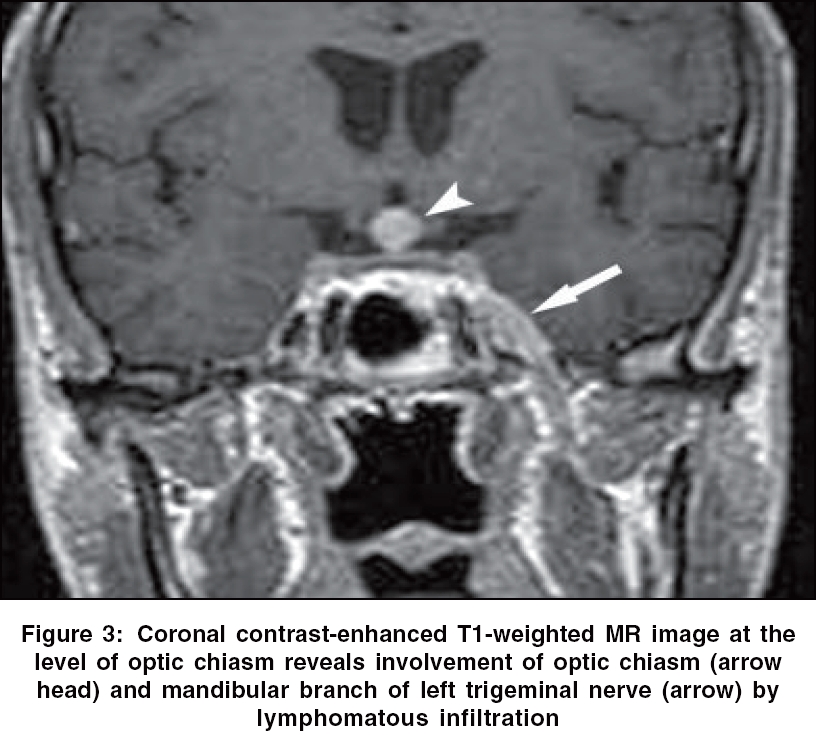
Neurology India, Vol. 54, No. 1, January-March, 2006, pp. 112-113 Neuroimage Cranial nerve lymphomatosis Kocaoglu M, Bulakbasi N, Bozlar U Department of Radiology, Gulhane Military Medical School, Etlik, Ankara Code Number: ni06038 Neurolymphomatosis is an extremely rare neurologic manifestation of systemic lymphoma in which B-cell nonHodgkin′s lymphoma a much more common cause. Neurolymphomatosis must be differentiated from more frequent neurologic manifestations of lymphoma, including peripheral nerves compression by enlarged lymph nodes, radiation plexopathy, herpes zoster infection, and lymphoma-associated vasculitis.[1] A nerve biopsy may show false negative results because of patchy nature of lymphomatous lesion;[2] therefore, sometimes diagnosis is made only at postmortem examination.[3] A 21-year-old male with diffuse large B-cell lymphoma, presented with the complaints of facial paralysis, dysphagia, and paraesthesia in both legs after the sixth course of CHOP regimen (cyclophosphamide, doxorubicin, vincristine and prednisone). Magnetic resonance (MR) imaging of head was performed with a 1.5 Tesla scanner. Axial and coronal thin section postcontrast T1 weighted MR images (TR: 600, TE: 16) revealed mass lesion in Meckel′s cave and optic chiasm and thickening of left abducens nerve, bilateral oculamotor nerves and mandibular branch of left trigeminal nerve consistent with lymphomatous infiltration. All lesions were contrast-enhanced following intravenous administration of paramagnetic contrast media. [Figure - 1][Figure - 2][Figure - 3] Several diagnostic procedures including electromyography, scintigraphy with Ga-67, computed tomography, and MR imaging may help in the diagnosis of neurolymphomatosis. MR imaging is a sensitive diagnostic tool that may demonstrate thickening, increased T2 signal and enhancement of the effected nerves on postcontrast T1-weighted scans and may help to identify potential sites for biopsy.[4] References
Copyright 2006 - Neurology India The following images related to this document are available:Photo images[ni06038f3.jpg] [ni06038f2.jpg] [ni06038f1.jpg] |
| |||||||||
