 |
| About Bioline | All Journals | Testimonials | Membership | News |
|
||||||
|
||||||
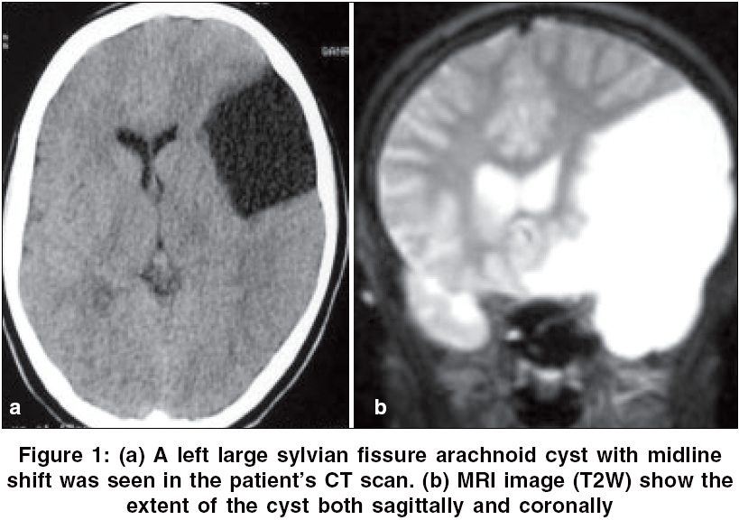
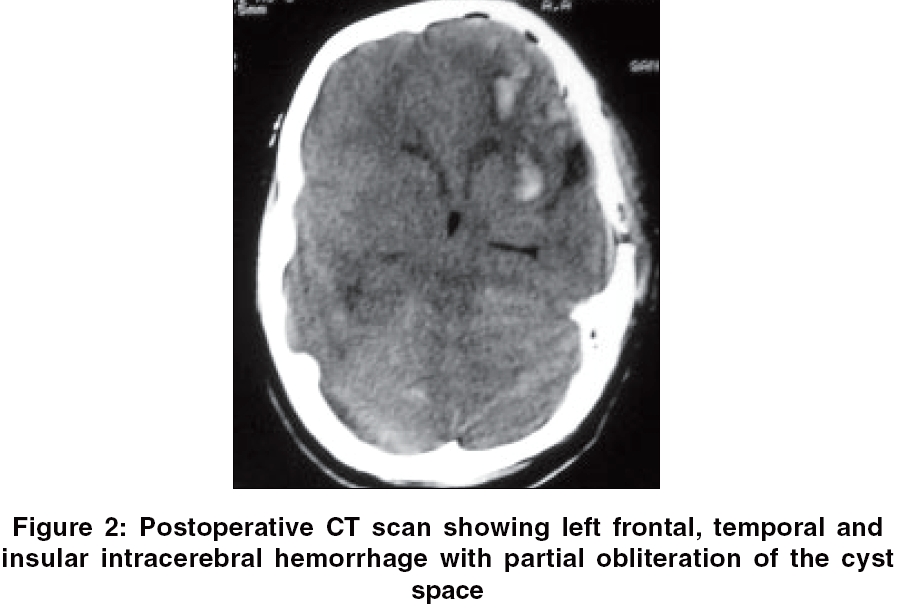
Neurology India, Vol. 54, No. 3, July-September, 2006, pp. 320-321 Letter To Editor Intraparenchymal hemorrhage after surgical decompression of a Sylvian fissure arachnoid cyst Esmaeeli Babak, Eftekhar Behzad Department of Neurosurgery, Shahrood Date of Acceptance: 21-Aug-2006 Code Number: ni06110 Sir, The management of the sylvian arachnoid cysts is still controversial.[1] Both direct opening of the cyst into the subarachnoid space and ′indirect′ surgical procedures (cysto-peritoneal shunting) are associated with complications. One of the rare complications after rapid decompression of the arachnoid cysts is hemorrhage in surrounding brain. We describe a case of intraparenchymal hemorrhage as a rare complication after surgical decompression of a sylvian fissure arachnoid cyst. A 15-year-old right-handed female presented with intractable generalized headache. Medical treatment for headache had failed. No neurological deficit or papilledema was seen in her neurological examination. In her preoperative CT scan and MRI, a left large sylvian fissure arachnoid cyst with midline shift was seen [Figure - 1]. Considering the mass effect, midline shift and intractable headache unresponsive to the medical management, the patient was admitted to the hospital for surgical decompression. After left-sided frontotememporal craniotomy, dura was opened and lateral wall of the cyst resected. A large draining vein on lateral wall of the cyst was preserved. The arachnoid cyst was opened to the basal cisterns uneventfully. After surgery, the patient opened her eyes and obeyed command. An hour later, a right-sided convulsive epilepsy unresponsive to antiepileptics occurred. Anesthetic dose of thiopental and mechanical ventilation began and an emergent computed tomography was performed. In the postoperative CT scan, left frontal, temporal and insular intracerebral hemorrhage with partial obliteration of the cyst space was seen [Figure - 2]. Unfortunately, the patient died on the seventh day after surgery with multiorgan failure. We could not do autopsy on the patient. Intraparenchymal hemorrhage in the underlying brain after decompression of the arachnoid cysts is an uncommon complication.[2] A case of brain stem hemorrhage after decompression of a sylvian fissure arachnoid cyst has been reported.[3] Intracerebral hemorrhage after rapid decompression of chronic subdural hematomas is well known and hyperperfusion of underlying brain after surgical decompression has been documented.[4],[5] Sgouros and Chapman have studied three children with middle fossa arachnoid cysts, presenting with nonspecific symptoms and otherwise well, before and after surgery with magnetic resonance and 99Tc-examethylpropyleneamineoxime single photon emission computerized tomography scans and shown that middle fossa arachnoid cysts may cause global impairment of brain function by interfering with its blood supply.[6] Although the pathophysiology of this complication is unclear, it might be due to re-perfusion injury, implying that there was raised intracranial pressure before cyst drainage. Other possible pathogenetic mechanisms of this complication are abrupt change in blood circulation, faulty autoregulation and brain decompression as the cause of superficial veins distortion. If this is the case, more gradual decompression of huge sylvian fissure arachnoid cysts using indirect surgical approaches with programmable shunts or more conservative procedures like simple tapping[1] may theoretically decrease the incidence of such rare complications in similar cases. References
Copyright 2006 - Neurology India The following images related to this document are available:Photo images[ni06110f1.jpg] [ni06110f2.jpg] |
| |||||||||
