 |
| About Bioline | All Journals | Testimonials | Membership | News |
|
||||||
|
||||||
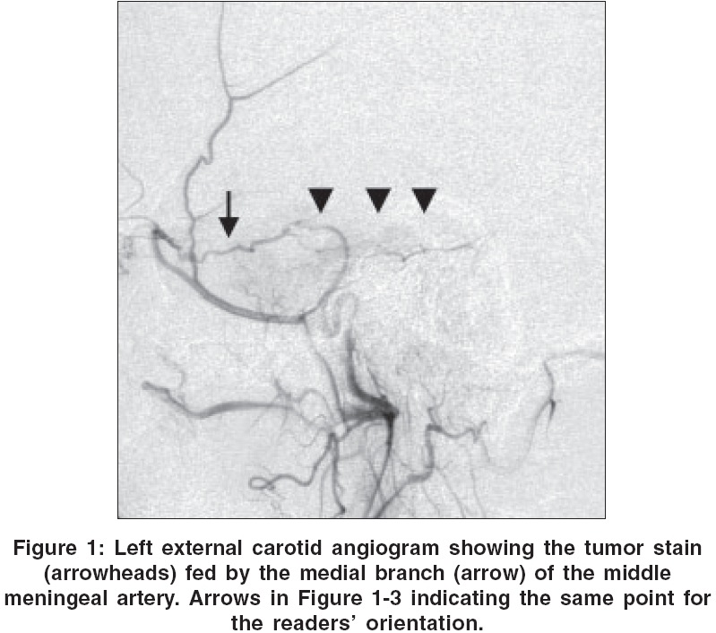
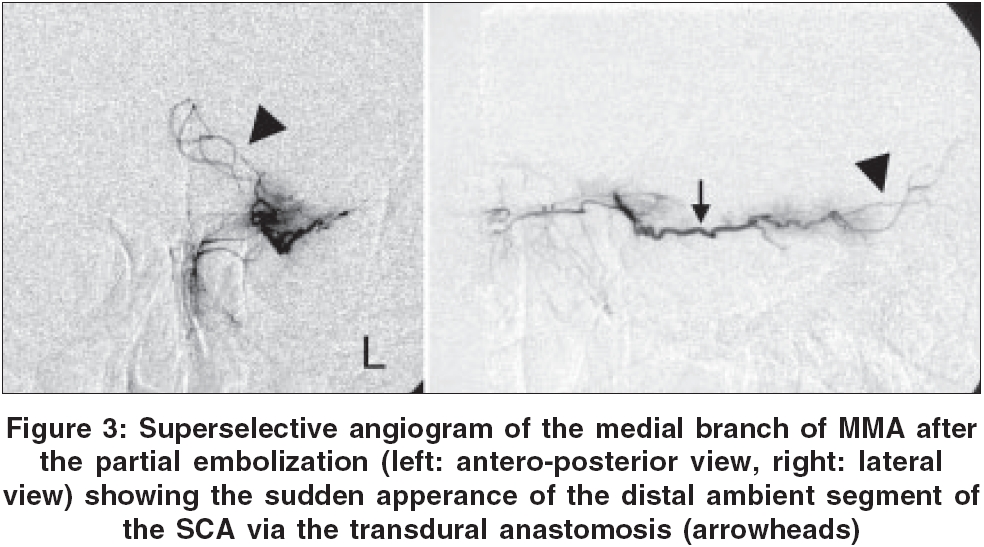
Neurology India, Vol. 54, No. 3, July-September, 2006, pp. 328 Neuroimage Sudden appearance of transdural anastomosis from middle meningeal artery to superior cerebellar artery during preoperative embolization of meningioma Ohata Kenji, Nishio Akimasa, Takami Toshihiro, Goto Takeo Department of Neurosurgery, Osaka City University Graduate School of Medicine, Osaka Date of Acceptance: 05-Feb-2006 Code Number: ni06116 We describe a rare transdural anastomosis to the superior cerebellar artery (SCA) in a 57-year-old man with cavernous sinus meningioma. This tumor had the feeders from the meningohypophyseal trunk (MHT) and the middle meningeal artery (MMA) on the lesion side [Figure - 1]. Preoperative embolization was performed with polyvinyl alcohol particles (PVA) of 45-150 mm in size via MHT, resulting in the subtotal embolization of this feeder. Subsequent superselective angiographies through the medial branch of the MMA confirmed the tumor stain without any pial supply [Figure - 2]. Unexpectedly, however, serial angiography during step-by-step embolization through this branch revealed the sudden appearance of the distal ambient segment of the left SCA via transdural anastomosis [Figure - 3]. Immediate discontinuance of the procedure could successfully prevent the embolic stroke. As far as the literature is concerned, we could find the brief description about this transdural anastomosis.[1] Connections of the branches of the MMA with the branches of other meningeal vessels or with the branches of the external and internal carotid arteries were proposed. The mechanism of development and the rate of establishment of such a pattern of transdural anastomosis are poorly understood. In such a situation, the awareness of the possible transdural anastomosis is important since the patient can present with an embolic stroke following embolization. References
Copyright 2006 - Neurology India The following images related to this document are available:Photo images[ni06116f2.jpg] [ni06116f1.jpg] [ni06116f3.jpg] |
| |||||||||
