 |
| About Bioline | All Journals | Testimonials | Membership | News |
|
||||||
|
||||||
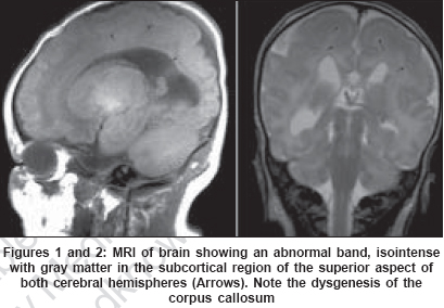
Neurology India, Vol. 55, No. 1, January-March, 2007, pp. 93 Neuroimage Band heterotopia in Zellweger syndrome (cerebro-hepato-renal syndrome) Young Sandra, Rabi Yacov, Lodha AbhayK Department of Pediatrics, Division of Neonatology, Foothills Medical Centre, Alberta Children Hospital, Institute of Maternal Child Health, University of Calgary/Calgary Health Region, Calgary, Alberta T2N2T9 Date of Acceptance: 27-Aug-2006 Code Number: ni07035 Zellweger syndrome (cerebro-hepato-renal syndrome) is associated with generalized hypotonia, high forehead with flattened facies, hepatomegaly and talipes equinovarus. This pattern of malformations was first recognized in 1964 by Bowen and Smith.[1] Zellweger syndrome is an autosomal recessive genetic disorder that is associated with multiple biochemical markers of peroxisomal dysfunction.[2] A full term, intrauterine growth restricted, male neonate was born to a 32-year-old, gravida 3, para 1 mother via spontaneous vaginal delivery. Polyhydramnios was noted in the pregnancy. Fetal ultrasonography demonstrated bilaterally enlarged ventricles in the brain and talipes equinovarus at 29 weeks of gestation. Apgar scores were 6 and 8 at 1 and 5min respectively. This patient presented with seizures at the time of birth and had classical findings suggestive of Zellweger syndrome including generalized hypotonia, large anterior fontanelle (6 x 6 cm), flat occiput, high forehead with shallow supraorbital ridges, mild micrognathia, weak suck, weak cry, single transverse palmar crease (simian crease), contractures at elbows and knees, short humeri, hepatomegaly, patent ductus arteriosus and ventricular septal defect and cryptorchidism. Zellweger syndrome is one of a number of peroxisome biogenesis disorders that can manifest as an absence or reduction in the number of peroxisomes in tissues as well as multiple enzymes abnormalities. Survivors have severe mental retardation and epileptic disorders.[2] Zellweger syndrome is associated with abnormal cortical gyral patterns, impaired myelination and cerebral periventricular pseudocysts.[3] In this infant, MRI of the brain showed an abnormal band that was isointense with gray matter in the subcortical region of the superior aspect of both cerebral hemispheres. Dysgenesis of the corpus callosum was also noted [Figure - 1 and 2]. References
Copyright 2007 - Neurology India The following images related to this document are available:Photo images[ni07035f1and2.jpg] |
| |||||||||
