 |
| About Bioline | All Journals | Testimonials | Membership | News |
|
||||||
|
||||||
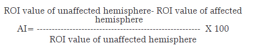
Neurology India, Vol. 56, No. 1, January-March, 2008, pp. 17-21 Original Article Pattern of cerebellar perfusion on single photon emission computed tomography in subcortical hematoma: A clinical and computed tomography correlation Kalita Jayantee, Misra UshaK, Ranjan Prasen, Pradhan PK Department of Neurology, Sanjay Gandhi Postgraduate Institute of Medical Sciences, Lucknow, Uttar Pradesh Date of Acceptance: 16-Jan-2008 Code Number: ni08004 Abstract Background: There is paucity of studies evaluating the role of asymmetry index (AI) on single photon emission computed tomography (SPECT) studies in patients with intracerebral hemorrhage (ICH).Aim: To evaluate cerebellar perfusion in ICH employing SPECT study and correlate with clinical and CT scan findings. Setting and Design: Tertiary care teaching hospital. Materials and Methods: A total of 29 patients with ICH were subjected to neurological examination including Glasgow Coma Scale (GCS) and Canadian Neurological Stroke Scale (CNS). Clinical features of raised intracranial pressure and herniation were noted. On CT scan, ICH location, volume, ventricular extension and midline (ML) shift were noted. On SPECT, cerebral and cerebellar perfusion was measured semiquantitatively and AI calculated. Outcome was defined at 3 months into poor and good. Results: Fourteen patients had putaminal and 15 thalamic hemorrhages. Their mean age was 59 years. The mean GCS score was 10 and CNS score 2.8. Hematoma was large in five, medium in 16 and small in eight patients. ML shift was present in 15 and hematoma extended to ventricule in 16 patients. On SPECT, cerebellar AI significantly related to ML shift but not with size of hematoma. AI was low in patients with ML shift. Outcome was related to GCS score, ML shift, size of hematoma and cerebellar AI. Conclusion: In acute stage of ICH, cerebellar AI is lower in patients with more severe stroke having ML shift. Keywords: Cerebellar diaschisis, computed tomography, hematoma, intracerebral hemorrhage, outcome, putaminal, SPECT, thalamic Intracerebral hemorrhage (ICH) is the commonest neurological emergency occurring in 10% patients with stroke. There is acute rise of intracranial pressure, and its severity depends on the size and location of hematoma. Basal ganglia and thalamic hematoma constitute majority of spontaneous ICH. Most patients with medium- and large-size hematoma are comatose and various outcome predictors such as size, location, ventricular extension, pupillary asymmetry, severity of stroke and Glasgow Coma scale (GCS) score have been reported. [1],[2],[3] With the availability of single photon emission computed tomography (SPECT) and PET, perfusion and cerebral metabolism, respectively, have been studied and their pattern and role in predicting outcome of stroke have also been reported. [4],[5] However, these studies are carried out mostly in patients with cerebral infarction. Role of cross-cerebellar diaschisis in supratentorial infarction has been highlighted and is attributed to interruption of the cerebropontocerebellar pathway that causes deafferentation and transneural metabolic depression of the contralateral cerebellum. [6],[7] Basal ganglia and thalamic hemorrhages may also disrupt the internal capsule. Thalamus being a major relay center for cerebellum, cross-cerebellar diaschisis (CCD) may also occur in these hematomas. The role of CCD in predicting outcome of stroke is controversial. [4],[8] In the available literature, there is paucity of reports describing pattern of cerebellar perfusion employing SPECT studies in basal ganglia and thalamic hematoma and their role in predicting outcome. In this communication, we report the pattern of cerebellar perfusion in subcortical hematoma in acute stage and its relation with clinical and CT scan changes. Materials and Methods Subjects A total of 29 patients with CT-proven thalamic and putaminal ICH were included in this study. Patients were admitted within 6 days of stroke and SPECT study was carried out within 48 h of admission. A detailed clinical evaluation was carried out. Depth of coma was assessed by GCS, severity of stroke by Canadian Neurological Scale (CNS) [9] and muscle power was graded into 0-5 as per Medical Research Council Scale. Muscle tone and tendon reflexes were also recorded. Sensations and cerebellar signs were evaluated only in those patients who could co-operate for these tests. Neurological signs of pyramidal dysfunction on the non-hemiplegic side as evidenced by increased reflex, tone or extensor plantar and pupillary asymmetry were noted. Cranial CT scan was carried out within 1 h of admission if not done earlier. Location of hematoma, ML shift, ventricular extension and size of hematoma were noted. Size of hematoma was categorized into small (< 20 ml), medium (20-40 ml) and large (>40 ml). None of these patients had infarction or hemorrhage in vertebrobasilar territory or any other supratentorial location except thalamic or putaminal ICH. None of these patients had a past history of stroke. Patients were managed in neurology ward and treated conservatively. Surgical evacuation or external ventricular drainage was not performed in these patients. The outcome of the patients were evaluated after 3 months on the basis of Barthel index (BI) score into poor (BI < 12) and good (BI > 12). [3] SPECT study SPECT study was carried out using 99m Tc ECD (ethylene cysteine dimer) employing a dual-headed gamma camera (DST XL; SMV, France) interfaced with a dedicated IBM computer system. Sixty-four projections were acquired with each projection for 40 s and the matrix size 128 x 128. A low-energy, high-resolution collimator was used. The transaxial sections were reconstructed by means of filtered back projection with a Butterworth filter with the order of 10 and cut-off frequency of 0.5. On average, 20 SPECT image planes, 3.9-mm thick, were required to image the entire brain. Axial sections were obtained parallel to orbitomeatal line and coronal and sagittal images were reconstructed. The SPECT images were analyzed semiquantitatively. A 10 x 10 mm region of interest (ROI) was placed on frontal, parietal, occipital, basal ganglia and the middle of ipsilateral and contralateral cerebellar hemisphere on the identical region. Total counts of ROI were obtained and asymmetry index (AI) was calculated as follows: [10]
Statistical analysis The correlation of AI with various clinical (GCS, CNS, pyramidal sign on non-hemiplegic side, pupillary asymmetry) and radiological parameters (size, ML shift and ventricular extension) were evaluated by correlation matrix. For evaluating the outcome predictors at 3 months, various clinical, radiological and AI were evaluated employing univariate logistic regression analysis followed by multiple logistic regression analysis using spss version 12.0 software. For statistical analysis, various demographic, clinical and radiological parameters were categorized as follows: gender - male = 1, female = 2; tone and tendon reflex - hypo = 1, hyper = 2, normal = 3 and pyramidal sign on non-hemiplegic side, pupillary asymmetry, ML shift and ventricular extension - present = 1, absent = 2. The raw data of age, GCS, CNS and volume of hematoma were used. The perfusion of frontal, parietal, occipital, basal ganglia and cerebellum with respect to ipsilateral and contralateral to hematoma was compared by independent t -test. Results The results were based on 29 patients with putaminal (14) and thalamic (15) hemorrhages who were admitted within 6 days of stroke. Their age ranged between 35 and 71 years (mean 59), and six were females. All were right-handed and none had previous history of stroke. The presenting symptoms were headache and vomiting in 14 each, altered sensorium in 28 and hemiplegia in all. None had seizures before or during the hospital stay. Seventeen patients were known hypertensive and seven detected to be hypertensive after developing stroke. Thirteen patients had neck rigidity. Muscle power was grade 0 in 20, grade I-II in 8 and grade III in 1 patient. Tone on hemiplegic side was reduced in 19 and increased in 9 patients. Deep tendon reflexes were decreased in 17 and increased in 11 patients. Evidences of contralateral pyramidal sign were present in eight and pupillary asymmetry in two patients. The mean GCS score was 10 (5-14) and CNS score was 2.8 (2-8.5). Cerebellar sign could not be elicited in any patient as they had altered sensorium; however, at 3-month follow-up none had cerebellar sign on the contralateral side. The admission BI score was 0 in all except one patient who had score of 12.The hematomas were thalamic in 15 and putaminal in 14 patients, which was large in 5, medium in 16 and small in 8 patients. ML shift was present in 15 patients; and in 16 patients, hematoma extended to ventricular system. SPECT The degree of cerebellar hypoperfusion was calculated as AI in the cerebellar hemisphere. The AI in the patients was 11.61 ± 5.88 (mean ± SD). On visual analysis, cerebellar hypoperfusion was noted on the ipsilateral side in seven patients; one of them had large, four medium and two small hematoma. Four of these seven patients with ipsilateral cerebellar hypoperfusion had ML shift. The mean cerebellar AI in patients without ML shift was 14.39, whereas it was 9.66 in patients with ML shift. Cerebellar AI correlated with CNS score ( r = 0.37, P = 0.049) and ML shift ( r = -2.10, P = 0.046) but not with GCS score ( r = 0.21, P = 0.29), size of hematoma ( r = 0.26, P = 0.17; [Figure - 1]), intraventricular extension of hematoma ( r = 0.005, P = 0.98), pupillary asymmetry ( r = 0.12, P = 0.54), pyramidal sign on non-hemiplegic side ( r = 0.34, P = 0.07) and outcome ( r = 0.03, P = 0.11). AI was more marked in patients with ML shift and low CNS score [Table - 1]. The perfusion of ipsilateral frontal, parietal and occipital areas was reduced insignificantly and details are given in [Table - 2]. Size of hematoma correlated with ML shift ( r = 0.55, P = 0.002), basal ganglia AI ( r = 0.43, P = 0.02) and frontal AI ( r = 0.47, P = 0.01). The cerebellar perfusion with respect to hematoma volume is shown in [Figure - 2]. At 3 months, 10 patients had poor, 14 partial and 5 complete recovery on the basis of BI score. On univariate analysis, outcome was significantly related to pyramidal sign on non-hemiplegic side ( P = 0.03), ML shift ( P = 0.001), size of hematoma ( P = 0.001), GCS score ( P = 0.01) and cerebellar AI ( P = 0.039) but not to pupillary asymmetry ( P = 0.09), intraventricular extension ( P = 0.13) and CNS score ( P = 0.51). The details are summarized in [Table - 2]. On multiple logistic analysis, the best set of parameters related to outcome was ML shift and GCS score [Table - 3]. Discussion In our study, cerebellar AI was lower in patients with ML shift and was related to severity of stroke as assessed by CNS score and 3-month outcome. In a study on right putaminal hemorrhage, there was a negative correlation between the degree of extension of hematoma to the internal capsule and regional blood flow of right motor cortex and bilateral cerebellar hemispheres. There was also a negative correlation between the volume of hematoma and regional blood flow of the right motor cortex and cerebellar hemispheres. [11] In our study, size of hematoma was also associated with ipsilateral frontal hypoperfusion as assessed by AI. Lower cerebellar AI in patients with ML shift suggests hypoperfusion of cerebellar hemisphere ipsilateral to hematoma. ML shift due to putaminal or thalamic hemorrhage may result compression of contralateral crus cerebri due to tentorial herniation, which may result in global cerebellar hypoperfusion and reduced AI. If acute compression of crus cerebri results higher CCD, this may even result apparently ipsilateral cerebellar hypoperfusion, which was seen in seven patients in our study. Ipsilateral cerebellar hypoperfusion in earlier studies with thalamic or putaminal hemorrhage has not been reported. [6],[8],[12],[13] This may be due to majority of studies on CCD are done on infarcts with mild to moderate severity. [6],[8] Studies on hematoma were also included mostly milder cases and SPECT studies were performed after 3 weeks when mass effect usually subsides. [12],[13] In our study, most of the patients were having severe stroke as assessed by CNS score and SPECT were performed within 8 days of the stroke. Therefore, we feel that reduced AI may be a sign of bilateral cerebellar hypoperfusion suggesting mass effect, ML shift producing compression on other side of crus cerebri. In the study by Ogasawara et al ., ipsilateral cerebellar hypoperfusion was not noted in patients with large hematoma. However, SPECT study in these patients was carried out after 29 days of stroke. [13] Outcome at 3 months in our study was related to GCS score, pyramidal sign to non-hemiplegic side, ML shift, size of hematoma and cerebellar AI. There are conflicting reports regarding relationship of cerebellar AI and outcome of stroke. However, studies on putaminal and thalamic hemorrhages are very few. In an earlier study, positive correlation between grade of motor function in subacute stage and BI in chronic stage with CCD have been reported. [13] However, their patient population was different from ours as out of 33 patients, hematoma were stereotactically aspirated in 9 and through craniotomy in 16 patients; and SPECT studies were performed after 29 days of stroke. None of our patients had undergone surgical evacuation and SPECT was performed within 8 days of stroke. SPECT studies performed during acute stage of infarctions did not correlate CCD with BI, CNS score and outcome. [5],[8],[10],[14] This has been attributed to (1) evolving cerebral perfusion and metabolic disturbances during acute stroke may alter the CCD findings and (2) CCD may be an unsettled pathological findings just after the stroke, which can manifest itself as reversible damage and potential tissue recovery. [4],[5],[15],[16] Low GCS score, size of hematoma and ML shift have been reported to correlate with outcome of stroke in a number of earlier studies. [2],[3],[17],[18] From this study, it can be concluded that SPECT study within 8 days of putaminal and thalamic hematomas not only reveals contralateral cerebellar hypoperfusion but can also ipsilateral cerebellar hypoperfusion. Cerebellar AI within one week of ICH is lower in more severe stroke as evidenced by CNS score and ML shift. Acknowledgement We thank Mr. Rakesh Kumar Nigam for secretarial help.References
Copyright 2008 - Neurology India The following images related to this document are available:Photo images[ni08004f2.jpg] [ni08004t1.jpg] [ni08004t3.jpg] [ni08004t2.jpg] [ni08004f1.jpg] |
| |||||||||

![[Figure - 1]](/showimage?ni/photo/ni08004f1.jpg)
![[Table - 1]](/showimage?ni/photo/ni08004t1.jpg)
![[Table - 2]](/showimage?ni/photo/ni08004t2.jpg)
![[Figure - 2]](/showimage?ni/photo/ni08004f2.jpg)
![[Table - 3]](/showimage?ni/photo/ni08004t3.jpg)