 |
| About Bioline | All Journals | Testimonials | Membership | News |
|
||||||
|
||||||
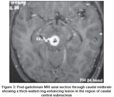
Neurology India, Vol. 56, No. 2, April-June, 2008, pp. 212-213 Letter To Editor Isolated bilateral ptosis as the presentation of midbrain tuberculoma Kumar Sudhir, Rajshekher Garikapati, Prabhakar Subhashini Department of Neurological Sciences, Apollo Hospitals, Hyderabad Code Number: ni08061 Sir, Isolated nuclear involvement of the oculomotor nerve is uncommon. Typical features of a nuclear third nerve lesion include unilateral third nerve palsy, bilateral superior rectus palsy and bilateral incomplete ptosis. [1] Here, we report a patient with bilateral incomplete ptosis without any other ocular or neurological signs and discuss the clinico-imaging correlation. A 14-year-old girl presented with headache and bilateral ptosis of two weeks duration. She had no diplopia or other neurological symptoms. On examination, she had bilateral symmetrical ptosis [Figure - 1], normal elevation of both eyeballs [Figure - 2] and normal pupillary size and reaction. Rest of the neurological examination was normal. The magnetic resonance imaging (MRI) brain revealed a thick-walled ring-enhancing lesion in the dorsal midbrain in the region of the oculomotor nucleus, possibly affecting the region of the caudal central (levator palpebrae superioris) subnucleus [Figure - 3]. Radiological features were suggestive of tuberculoma. She was empirically started on antituberculous treatment and steroids, with which she showed clinical improvement. Isolated bilateral ptosis has been previously reported in association with midbrain lesions due to subacute encephalitis [2] and midbrain hemorrhage. [3] It should be noted, however, that it is more common to find ptosis in association with upgaze paresis. This is because the unpaired superior rectus subnuclei are located medially in close association with the caudal central subnucleus (levator palpebrae subnucleus). This case is reported for its unique clinical presentation, which can be explained on the basis of lesion location on MRI. To the best of our knowledge, isolated bilateral ptosis due to midbrain tuberculoma has not been previously reported. References
Copyright 2008 - Neurology India The following images related to this document are available:Photo images[ni08061f1.jpg] [ni08061f2.jpg] [ni08061f3.jpg] |
| |||||||||
