 |
| About Bioline | All Journals | Testimonials | Membership | News |
|
||||||
|
||||||
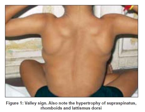
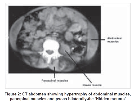
Neurology India, Vol. 56, No. 3, July-September, 2008, pp. 394 Neuroimage 'Hidden mounts' in Duchenne muscular dystrophy Suresh Chandran CJ Department of Neurology, Kerala Institute of Medical Sciences, Trivandrum, Kerala Correspondence Address:Kerala Institute of Medical Sciences, Trivandrum, Kerala drceejay@rediffmail.com Code Number: ni08096 A ten-year-old boy presented with inability to run, progressive difficulty in walking and weakness of proximal muscles of lower and upper limbs from the age of three years. By the time he presented to us, he was unable to stand even with support. Neurological examination revealed hypertrophy of supraspinatus, infraspinatus, deltoid, lattismus dorsi, rhomboids and calf muscles, bilaterally. ′Valley sign′ was present [Figure 1]. Serum CPK was 8000 U/L. EMG showed myopathic potentials. Polymerase chain reaction identified dystrophin gene deletion mutation and diagnosis of Duchenne muscular dystrophy (DMD) was confirmed. Abdominal CT cuts were taken to evaluate for abdominal muscles. CT abdomen showed hypertrophy of abdominal muscles, paraspinal muscles and psoas bilaterally-′hidden mounts′ [Figure 2]. Calf muscles are the commonest muscles to undergo hypertrophy in DMD, followed by infraspinatus. But various other muscle groups also show enlargement in DMD. Hypertrophy of abdominal and paraspinal muscles are probably overlooked. Our patient had hypertrophic abdominal muscles, paraspinal muscles and psoas bilaterally as demonstrated in the abdominal CT- the ′hidden mounts′. Valley sign is a specific sign of DMD with 90% sensitivity. It is due to the wasting of muscles in the posterior axillary fold (valley). On either side of this valley there are two mounts -hypertrophic infraspinatus inferomedially and deltoid hypertrophy superolaterally. The whole appearance is like a ′valley between two mounts′. [1] References
Copyright 2008 - Neurology India The following images related to this document are available:Photo images[ni08096f1.jpg] [ni08096f2.jpg] |
| |||||||||
