 |
| About Bioline | All Journals | Testimonials | Membership | News |
|
||||||
|
||||||
Neurology India, Vol. 57, No. 1, January-February, 2009, pp. 98 Letter To Editor Extradural thoracic spinal meningioma Bruno M. Santiago, Paula Rodeia, Manuel Cunha e Sá Department of Neurosurgery, Hospital Garcia de Orta, Av. Torrado da Silva, 2801-951, Almada, Portugal Correspondence Address: Department of Neurosurgery, Hospital Garcia de Orta, Av. Torrado da Silva, 2801-951, Almada, Portugal brunosantiago@sapo.pt Date of Acceptance: 26-Jan-2009
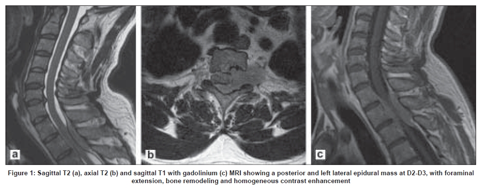
Code Number: ni09031 Sir, A 42-year-old male patient was admitted for the investigation of an insidious onset Grade 4 paraparesis progressing over a period of one month. Magnetic resonance imaging (MRI) revealed a posterior and left lateral epidural mass at D2-D3, with foraminal extension and bone remodeling of the left posterior segment of the D3 vertebral body. The lesion was isointense to the spinal cord both on T1 and T2 sequences and with an intense and homogeneous gadolinium uptake [Figure 1 a-c]. A D2-D3 laminectomy was performed and a totally extradural tumor mass, very adherent to the dura was removed. Neuropathology reported a typical psammomatous meningioma, the most common histological subtype of spinal meningiomas. Meningiomas account for 25% of all intraspinal neoplasms and are the second most common primary intraspinal tumor. Exclusively epidural meningiomas are very rare, accounting for 2,7-3,5% of spinal meningiomas, but in some large series they were not reported. [1],[2] Their origin is probably in the ectopic extradural arachnoid cells. These tumors can be intracanalar with dural sac compression simulating a metastasis or hematological malignancy, or they can have a foraminal location or extension, making them difficult to distinguish from a schwannoma. The iso or hypointensity of this type of lesion on T2 MRI sequences contrasts with the T2 hyperintensity of most epidural tumors, except for lymphomas that can be hypointense in over 50% of cases. Spinal neurinomas can be exclusively extradural in 15% of cases, with a post-ganglionic origin and foraminal extension to the intraspinal compartment and epidural compression (Type IV spinal schwannomas). They tend to be hypointense to the spinal cord on T1 sequences while meningiomas are more frequently isointense. The clinical behavior of extradural meningioma seems to be no different from its intradural counterpart. Although these lesions are rare it is very important to be aware of them, given their different prognosis and rate of surgical cure when compared with the more frequent tumors of this spinal compartment. References
Copyright 2009 - Neurology India The following images related to this document are available:Photo images[ni09031f1.jpg] |
| |||||||||
