 |
| About Bioline | All Journals | Testimonials | Membership | News |
|
||||||
|
||||||
Neurology India, Vol. 57, No. 1, January-February, 2009, pp. 98-99 Letter To Editor Ruptured intracranial dermoid cyst Santosh P. V. Rai Department of Radiodiagnosis, KMC Hospital, Attavar, Mangalore - 575 001, Karnataka, India Correspondence Address:Department of Radiodiagnosis, KMC Hospital, Attavar, Mangalore - 575 001, Karnataka, India radiorai@gmail.com Date of Acceptance: 21-Jan-2009
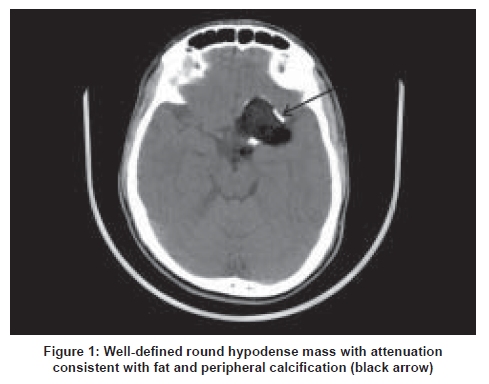
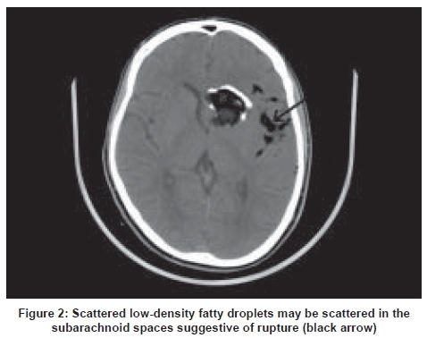
Code Number: ni09032 Sir Intra-axial dermoid cysts are rare intracranial lesions, more so in the pediatric age group. Dermoid cysts account for about 0.2 to1.8% of all intracranial tumors and are commonly located in the cisternal spaces, mainly in the cerebellopontine angle and parasellar cisterns. [1] Intracranial dermoid cysts are pathologically characterized by a thick, stratified squamous epithelium cyst wall containing dermal elements. [2] Rupture of dermoid cyst can cause granulomatous chemical meningitis that can result in recurrent symptoms, most commonly headache. Headache is often the presenting feature of ruptured intracranial dermoid. Rupture of dermoid cyst is unusual to present in older people. [3] Rupture of an intracranial dermoid produces a dramatic MR and CT appearance. [4] Computerized tomography (CT) scan typically shows a well-defined round hypodense mass lesion with attenuation consistent with fat and peripheral calcification [Figure - 1]. In case of ruptured dermoid cyst, CT scan shows low-density fatty droplets scattered throughout the ventricles and subarachnoid space [Figure - 2]. A fat-cerebrospinal fluid (CSF) level may also be seen. Dermoid cysts do not enhance on contrast administration. The presence of disseminated fat droplets in the subarachnoid space or ventricles on neuroimaging is diagnostic for a ruptured dermoid cyst. [5] A definitive diagnosis can be made by the characteristic features on CT scan [6] Magnetic resonance imaging typically demonstrates high signal intensities on T1 and variable signal intensities on T2. This is consistent with the lipid and cholesterol which typically collects within the dermoid cyst. When the cyst ruptures, high-signal droplets on T1 images may be seen scattered throughout the CSF. Sometimes a fat-CSF fluid level may also be seen. References
Copyright 2009 - Neurology India The following images related to this document are available:Photo images[ni09032f2.jpg] [ni09032f1.jpg] |
| |||||||||
