 |
| About Bioline | All Journals | Testimonials | Membership | News |
|
||||||
|
||||||
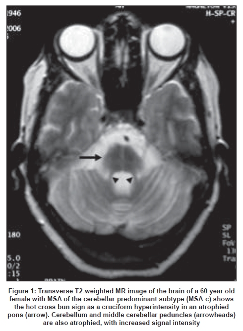
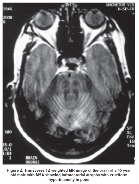
Neurology India, Vol. 57, No. 1, January-February, 2009, pp. 104-105 Neuroimage The hot cross bun sign A. Gulati, V. Virmani, P. Singh, N. Khandelwal Department of Radiodiagnosis and Imaging, Postgraduate Institute of Medical Education and Research, Sector 12, Chandigarh - 160 012, India Correspondence Address: Dr. Ajay Gulati, Department of Radiodiagnosis, PGIMER, Chandigarh, India. drajaygulati@rediffmail.com Date of Acceptance: 26-Jan-2009
Code Number: ni09035 Hot cross bun sign refers to the cruciform-shaped hyperintensity on T2W axial magnetic resonance images (MRI) in multisystem atrophy due to the selective loss of myelinated transverse pontocerebellar fibers and neurons in the pontine raphe and sparing of the pontine tegmentum and corticospinal tracts [Figure - 1],[Figure - 2]. [1] The name derives from a sweet spiced bun baked by the Christian church on the last Thursday before Easter and marked with a cross on the top, with the four quarters representing the four quarters of the year. Multisystem atrophy (MSA) is a sporadic progressive neurodegenerative disorder of adult onset, involving the basal ganglia and the olivopontocerebellar complex to varying degrees. It commonly presents with Parkinsonian symptoms and cerebellar ataxia and depending on which of these symptoms predominates, is classified as MSA-Parkinsonian predominant(MSA-P) and MSA-Cerebellar predominant (MSA-C) respectively. [2] MSA, and especially MSA-P, has to be differentiated from Parkinson's disease (PD) with which its clinical features overlap but treatment may differ. Certain findings on MRI may be a useful aid to clinical diagnosis. Atrophy of the putamen and brainstem and abnormal signal in the middle cerebral peduncle are found in a significant number of patients with MSA, while they are almost never found in PD patients. [3] Other findings like hypointensity of putaminal body, slit-like hyperintensity of the lateral putaminal border and atrophy of the cerebellar vermis or hemispheres may be found in both MSA and PD, but the findings are usually mild in PD and when moderate or severe point to the diagnosis of MSA. [3] Hot cross bun sign is a manifestation of pontine atrophy in MSA and is more commonly found in MSA-C. Infratentorial atrophy with hyperintense signal in the middle cerebral peduncle and the hot cross bun sign in the pons can provide helpful adjunct in making the diagnosis of MSA. References
Copyright 2009 - Neurology India The following images related to this document are available:Photo images[ni09035f1.jpg] [ni09035f2.jpg] |
| |||||||||
