 |
| About Bioline | All Journals | Testimonials | Membership | News |
|
||||||
|
||||||
Neurology India, Vol. 57, No. 2, March-April, 2009, pp. 143-150 Original Article Hyperdense middle cerebral artery sign in multidetector computed tomography: Definition, occurrence, and reliability analysis Kasim Abul-Kasim, Eufrozina Selariu, Marco Brizzi 1 , Jesper Petersson1 Faculty of Medicine, Section of Neuroradiology, Departments of Radiology and Correspondence Address: Dr. Kasim Abul-Kasim, Faculty of Medicine, University of Lund, Department of Radiology, Section of Neuroradiology, Malmö University Hospital, 205 02 Malmö, Sweden. kasim.abul-kasim@med.lu.se Date of Acceptance: 12-Mar-2009
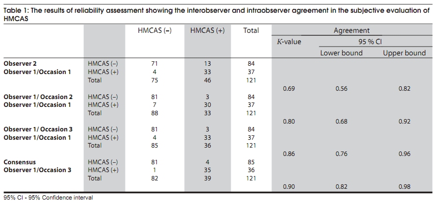
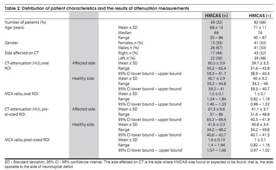
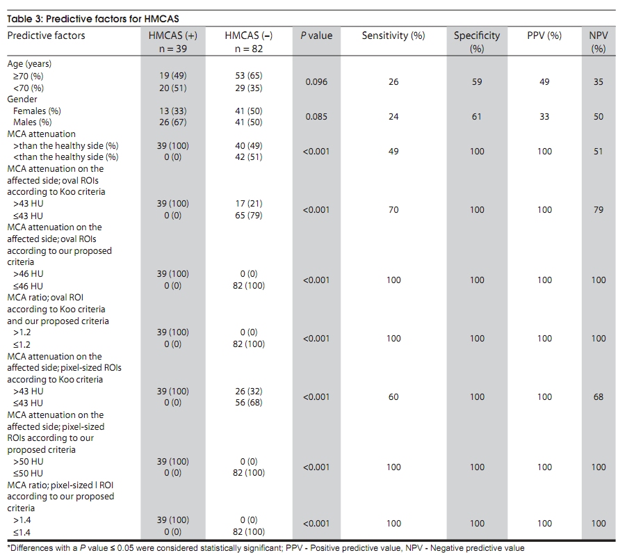
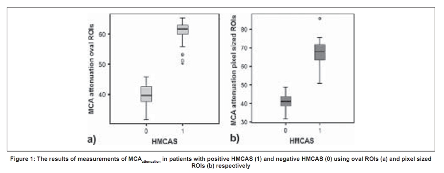
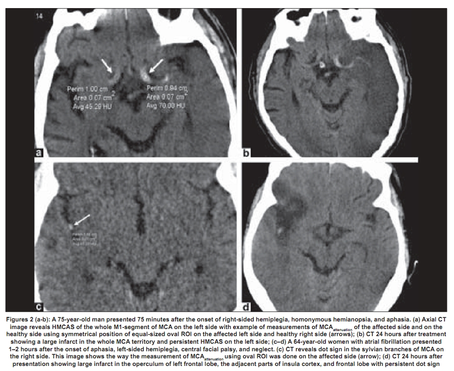
Code Number: ni09043 PMID: 19439843 DOI: 10.4103/0028-3886.51282 Abstract Background: The hyperdense middle cerebral artery sign (HMCAS) is one of the early changes seen on the computed tomography in acute ischemic stroke of MCA territory.Aims: To evaluate the reliability of subjective evaluation of HMCAS on CT performed at multidetector CT (MDCT) and evaluated in the Picture Archiving Communication Systems, to define objective criteria for HMCAS and to find out if there are any predictors for the occurrence of HMCAS. Materials and Methods: CTs of 121 consecutive patients (mean age of 70 years) treated with thrombolytic therapy were retrospectively evaluated by two neuroradiologists both subjectively and objectively with respect to HMCAS. Results: HMCAS was subjectively found in 32% of study population. The interobserver and intraobserver agreement were substantial (K value of 0.69 and 0.80, respectively) and increased to almost perfect (Kvalue of 0.86) when the reader provided with clinical information. The HMCAS was found twice as often in male patients. Patients with HMCAS were three years younger than those whose baseline CT did not show HMCAS. A 100% sensitivity achieved when objective criteria were defined as combination of MCA attenuation ≥ 46HU and MCA ratio > 1.2 (using oval ROIs) and MCA attenuation ≥ 50 HU and MCA ratio of > 1.4 (using pixel sized ROIs). Conclusion: Performing CT examinations on MDCT and assessment of the images in PACS might have contributed to improvement of the reliability of evaluating HMCAS on CT by enabling an objective evaluation of this sign with measurements of attenuation value in the course of MCA using oval or pixel sized ROIs as well as estimation of MCA ratio . Keywords: HMCAS, MCA attenuation, MCA ratio, MDCT, PACS, stroke Introduction Computed tomography (CT) and magnetic resonance imaging (MRI) play a central role in the management of acute stroke. CT perfusion (CTP), diffusion-weighted imaging (DWI), and perfusion-weighted imaging (PWI) may help to improve identification of patients suitable for treatment with tissue plasminogen activator (t-PA). However, MRI is less available than CT and often poses logistic difficulties when handling severely sick stroke patients. Because of better availability, ease of use, and short examination time, CT is still (and is likely to remain) the method of choice in the initial workup of stroke, [1] with the primary task to exclude intracranial hemorrhage. However, CT angiography (CTA) and CTP play an important role in the management of stroke in patients admitted beyond the therapeutic window in order to find out if there is any salvageable brain tissue and in the decision taking before intra-arterial thrombolysis therapy. Multimodal CT evaluation has been shown to improve detection rate and prediction of the final size of infarction compared with unenhanced CT, CTA, and CTP alone. [2] CTA demonstrates the anatomical details of the brain vasculature and is shown to be highly accurate in the identification of occlusions in the proximal large vessels in the circle of Willis, and in the rapid triage of patients to intra-arterial or intravenous (IV) thrombolytic therapy. [3] CTP can provide important prognostic information regarding final infarct size and clinical outcome in acute (< 6 hours post onset) stroke patients. [4] However, in centers and in countries where the availability of these advanced modalities is limited plain CT remains the method of choice in the initial workup of acute ischemic stroke. Early ischemic changes (EICs) on CT have been systematically reviewed [1] and are also to be looked for in the baseline CT. Loss of insular ribbon, obscuration of sylvian fissure, cortical sulcal effacement, obscuration of lentiform nucleus, loss of gray and white matter differentiation in the basal ganglia and focal hypoattenuation, all represent EICs. The hyperdense middle cerebral artery sign (HMCAS) is another important sign to be looked for on the baseline CT in acute ischemic stroke. HMCAS is a radiological finding first described in 1983, [2] and defined as increased attenuation of middle cerebral artery (MCA) indicating thromboembolic occlusion of the main trunk (M1-segment) of MCA. [5] Presence of HMCAS was proven to be associated with large infarcts and poor prognostic outcome [6],[7],[8],[9],[10] and thus represents important information for the treating clinician. Hyperdensity in the sylvian branches of MCA (M2-segment), known as 'dot sign', indicates a distal embolic occlusion and is associated with better prognosis compared with HMCAS of M1-segment. [11] HMCAS is a subjective finding and studies have previously been performed to establish objective features of HMCAS. Koo et al ., defined hyperdensity as absolute MCA attenuation > 43 Hounsfield units (HU) and MCA ratio > 1.2 (MCA attenuation of the affected side/MCA attenuation of the healthy side), [12] while Schuknecht et al ., found CT attenuation of 77-89HU in the vessels subjectively considered as hyperdense. [13] However, CT techniques have substantially improved and the validity of these findings needs to be reevaluated. The major aim of this study was to evaluate the interobserver and intraobserver agreement in the assessment of the subjective evaluation of HMCAS on the baseline nonenhanced brain CT of patients examined within three hours of onset of stroke in the MCA territory and subjected to thrombolytic therapy. The other aims of the study were to test the validity of criteria for objective features of HMCAS defined by Koo et al ., and Schuknecht et al ., as well as to find out if there is (are) any predictor for the occurrence of HMCAS. Materials and Methods The subjects of this retrospective study were all patients treated with IV recombinant t-PA between February 2004 and April 2008. A total 121 consecutive patients with stroke in the MCA territory were identified in the stroke register of this period and were included for the analysis. Fifty five percent patients were males and 45% were females with mean age of 70 ± 12 years (mean ± SD), median age of 72 years, and range of 30-87 years. All patients were examined with a baseline nonenhanced CT of the brain (from foramen magnum to vertex) using a multidetector CT (MDCT) (SOMATOM Sensation 16, Siemens AG, Forchheim, Germany) with a slice collimation of 0.75 mm and slice width of 4.5 mm. Reformatted axial images with soft tissue algorithm and with slice thickness of 4.5 and 0.75 mm were obtained. Furthermore, 3-mm thick reformatted coronal images were also obtained. All CTs were evaluated independently by two observers (two neuroradiologists) at two different occasions (with two weeks interval) with no knowledge of the symptom or the side affected. Images were evaluated only with regard to occurrence of HMCAS. At a third occasion, two weeks later, one observer provided with information about the side and the type of the neurological deficit. In cases of disagreement between observers 1 and 2, a joint evaluation by the two observers was performed to reach a consensus about the occurrence of HMCAS. The aim of the evaluation at the third occasion was to evaluate the degree of agreement between the results at this occasion and the results of the consensus reached at the joint evaluation panel. With the evaluation at this occasion we also sought to explore the impact of the successively increasing experience of the evaluating radiologist on the assessment of the occurrence of HMCAS. The evaluation of all images in our study was performed in picture archiving and communication systems (PACS). For objective evaluation of HMCAS, CT attenuation in HU was measured on axial images by two different methods. The evaluating radiologist was blinded to the symptom or the side affected as well as to the results of the subjective evaluation of HMCAS. As performed by Koo et al ., the MCA attenuation was first measured by placing oval or elliptical region of interest (ROI) over MCA on both sides. The size of the ROIs was dependant on the size of the vessel segment met on different axial cuts with the aim to place equal-sized-mirror ROIs on either side and to only include the vessel lumen to avoid influence from the surrounding parenchyma or cerebrospinal fluid. The area of ROIs ranges between 0.05-0.07cm 2 in cases of HMCAS involving the main trunk of MCA and 0.01-0.03cm 2 in cases of dot sign. Because MCA may be met at different cuts, MCA attenuation in five ROIs on the affected side and five ROIs on the healthy side were measured and the mean value was calculated for each side separately in every individual examination. The MCA ratio for every individual examination was also calculated. Furthermore, MCA attenuation was measured using five ROIs of one pixel size (as performed by Schuknecht et al .) at five different pixels on the affected side and at five different pixels on the healthy side. MCA ratio for every individual examination was also calculated. The affected side was defined as the side where HMCAS signs were subjectively seen or expected to be seen which is contralateral to the side of neurological deficit. All patients treated with t-PA were subjected to CT control 24 hours after the initiation of thrombolysis therapy for evaluation of the occurrence of infarction in the MCA-territory, evaluation of whether HMCAS disappeared or persisted after treatment in patients whose initial CT showed HMCAS, estimation of the infarct volume, and evaluation with regard to Alberta stroke program early CT score (ASPECTS). [14] On deterioration, CT was performed earlier to rule out cerebral hemorrhage. Statistical analysis Statistical analysis was performed with SPSS 15 (originally Statistical Package for the Social Sciences). The degree of interobserver and intraobserver agreement in the subjective evaluation regarding the occurrence of HMCAS was evaluated by cross tabulation and calculation of kappa (K values). The interpretation of kappa values was done according to the one proposed by Landis and Kock. [15] A kappa of one indicates complete agreement of all observers in all cases whereas a kappa of zero indicates that any observed agreement is attributed to chance. To explore the probability of different factors (age, gender, MCA attenuation , and MCA ratio ) to predict the occurrence of HMCAS chi-square test was performed. SPSS automatically computes Fisher's exact test in addition to chi-square test when the table has a cell with an expected frequency of less than five. Differences with a P value ≤ 0.05 were considered statistically significant. Sensitivity, specificity, positive predictive value (PPV), and negative predictive value (NPV) of every analyzed predictor have been calculated. Results The joint evaluation panel was agreed that 39 patients (32%) showed radiological evidence of thromboembolism in the MCA territory; 32 patients with HMCAS; and 7 patients with dot sign. Seventeen patients (44%) showed HMCAS on the right side. The results of reliability analysis (interobserver and intraobserver agreement) in the subjective evaluation of HMCAS and dot sign on baseline CT are shown in [Table - 1]. Observer 1 and observer 2 were agreed about the occurrence of HMCAS in 104 patients while observer 1 was agreed about the occurrence of HMCAS in 111 patients at two different occasions which resulted in substantial interobserver and intraobserver agreement (K value 0.69 and 0.80, respectively). When the reader was provided with information about the symptoms and the side of neurological deficit (observer 1 at the third occasion), the degree of agreement increased to almost perfect compared with the consensus reached at the joint evaluation (K value 0.90). In all cases with HMCAS the side considered revealing this sign was contralateral to the side of neurological deficit. The different characteristics of patients whose baseline CT subjectively showed HMCAS and those whose baseline CT did not show HMCAS are shown in [Table - 2] (labeled as HMCAS (+) and HMCAS (-), respectively). Patients whose baseline CT subjectively did not show HMCAS were three years older than those with HMCAS (71 ± 11 years (mean ± SD) and 68 ± 13 years (mean ± SD), respectively). In the patient group with negative HMCAS 65% were ≥70 years compared with 49% in the patient group with positive HMCAS. However, this difference was not statistically significant ( P = 0.096), [Table - 3]. The occurrence of HMCAS was unrelated to gender, although 67% of patients with positive HMCAS were males compared with 50% in patients with negative HMCAS ( P = 0.085), [Table - 3]. The results of measurements of MCA attenuation and MCA ratio obtained using oval ROIs and pixel-sized ROIs are shown in [Table - 2] and [Figure - 1]. The mean value of MCA attenuation of the affected side was 1.5 times higher than that of the healthy side (MCA ratio ) when the measurements were done with oval ROIs compared with 1.6 times when the measurements were done with pixel-sized ROIs. The MCA attenuation was higher on the affected side in all patients who subjectively showed HMCAS ( n = 39) regardless of the type of ROIs used for the measurements. The occurrence of HMCAS was related to objective criteria proposed by Koo et al . [9] All patients who subjectively showed HMCAS had MCA attenuation on the affected side higher than 43 HU and MCA ratio higher than 1.2 ( P = < 0.001), [Table - 3]. However, 17 patients (21%) who were subjectively not considered to have HMCAS ( n = 82) had MCA attenuation on the affected side higher than 43 HU (objectively false positive HMCAS) but none of them had MCA ratio higher than 1.2. Increasing the limit of MCA attenuation from 43 HU to 46 HU was found to increase the sensitivity and the NPV of the objective criteria proposed by Koo et al ., to 100%, [Table - 3]. Using pixel-sized ROIs, all patients who subjectively showed HMCAS had MCA attenuation of> 50HU and MCA ratio > 1.4 compared with those whose baseline CT did not show HMCAS ( P = < 0.001), [Table - 3]. The occurrence of MCA stroke in our study was confirmed by the follow-up CT examination performed 24 hours after thrombolysis therapy. All patients with HMCAS sustained cerebral infarct in the MCA territory. At CT control 24 hours after treatment, HMCAS remained positive in 17 (44%) patients, moved from M1 to M2 and M3 segments of MCA (conversion to dot sign) in 8 (20%) patients, and disappeared in the remaining 14 (36%) patients. Patients who had positive HMCAS or those with HMCAS being converted to dot sign after treatment sustained larger infarcts compared with patients whose HMCAS disappeared with average ASPECTS of 3.6 and 6, respectively, (infarction of the whole MCA territory: ASPECTS 0 whereas normal CT: ASPECTS 10. [14] ) [Figure - 2] shows images of the baseline nonenhanced CT of two different patients with HMCAS and dot sign, the way the objective evaluation using oval ROIs was performed, and images of the follow-up CT performed 24 hours after treatment with thrombolysis. Discussion There is an ongoing controversy on clinical significance, reliability, and prognostic value of EICs on CT with regard to the attenuation changes in different parts of the MCA territory. [16],[17] However, HMCAS is a radiological finding which is distinct and easy to identify. The specificity of HMCAS for MCA thromboembolic occlusion may be as high as 100%, while the sensitivity is rather low. [6],[18] The most common sources for false positive HMCAS are calcification in the wall of MCA and increased hematocrit.[6],[19] Bromide therapy and use of cocaine have been reported to increase vascular density in absence of thrombosis. [20],[21] The prevalence of HMCAS varies widely depending on the population studied. Among patients with infarction in the MCA territory the prevalence of HMCAS were reported to occur in 30% of cases. [19] The reliability of CT in the assessment of EICs was studied before the era of MDCT.[22] In that study the interobserver agreement was moderate to substantial for HMCAS on right and left hemisphere, respectively (K value of 0.62 and 0.57, respectively). The higher degree of agreement in our study is probably due to improvement of CT technology. CT examinations in our study were performed on a MDCT with a slice collimation of 0.75 mm, which allows obtaining images with a slice thickness of 0.75 mm as well as the possibility to obtain reformatted images in the coronal plane. Furthermore, the evaluation of all images in our study was performed in PACS which allows changing window center and window width (brightness and contrast) of the evaluated image. Other applications such as the ability to pan and zoom images and to measure the attenuation have helped to improve the assessment of the MCA density. The radiation dose is a limiting issue in the radiological workup including CT and other modalities using the ionizing radiation. However, modern CT scanner enables angular tube current modulation as well as modulation in z-axis, [23] with the aim to automatically adapt the tube current to the patient's anatomic configuration and size. PACS also provides opportunity to objectively assess the HMCAS by enabling measurement of the MCA attenuation. Although the degree of interobserver agreement in our study was already high (K value of 0.79) when the observers were blinded to the clinical information, the degree of intraobserver reliability markedly increased when the evaluation was performed with knowledge of symptoms and the side affected. This emphasizes the importance of providing the neuroradiologist with clinical information when evaluating the images of the baseline CT in patients with stroke prior to treatment with t-PA. The increased intraobserver agreement between occasions 2 and 3 might reflect an increased experience of the evaluating neuroradiologist, gained from repeated assessments, in the detection of this sign and emphasizes the importance of reader's experience in the evaluation of HMCAS and probably other signs of EICs. However, an interval of two weeks between occasions 2 and 3 is rather short and there is a small but existing possibility that the reader remembered the findings in some of the evaluated images which may subsequently have contributed to the increasing agreement between the second and the third occasion. The objective evaluation of HMCAS was shown to be dependent on the method of measurement of the CT attenuation. Performing the measurements by using oval ROIs showed higher mean value of MCA attenuation of the affected side in our study (60.5 ± 3.9 (mean ± SD), 95% CI 59.3-61.7) compared with the results obtained by Koo et al ., (54 HU (mean), 99% CI 46.7-61.2). The occurrence of atherosclerosis and vessel wall calcifications might have contributed to this difference as the mean age in our study was almost 20 years higher than that of patients included in the reports by Koo et al ., (68 ± 13 years (mean ± SD) versus 49 ± 22 years (mean ± SD)). Although application of Koo criteria (MCA attenuation > 43) in measurements of MCA attenuation using pixel-sized ROIs showed statistically significant difference between the MCA attenuation of the affected side and the healthy side ( P = < 0.001), 32% of patients ( n = 26) who subjectively did not show HMCAS had MCA attenuation > 43, [Table - 3]. Furthermore, Schuknecht et al ., did not report any data on MCA ratio and their measurements of CT attenuation included different intracranial and extracranial vessels. With this background we sought to define a cut-off value for MCA attenuation and MCA ratio when such measurements were performed using ROIs of one pixel size. Since all patients who subjectively considered having HMCAS had MCA attenuation > 50 HU and MCA ratio > 1.4, these values were considered as defining the limit between the positive and negative HMCAS. Although conventional angiography remains gold standard to confirm the presence of thrombosis in the MCA, CTA as well as magnetic resonance angiography (MRA) has been increasingly used for this purpose. None of these modalities were included a routine in the workup of stroke in our study which was probably one of the limitations of this study. However, the CT control 24 hours after treatment showed that HMCAS disappeared or moved from M1 to M2 and M3 segments in 22 patients, which confirms that the increased attenuation in the MCA detected at the baseline nonenhanced CT represented HMCAS attributed to thromboembolism of MCA rather than vessel wall calcifications which should have persisted. The remaining 17 patients with persistent HMCAS after treatment developed large infarcts in the MCA territory with average ASPECTS of 3.6. Unilaterality of HMCAS and the compatibility of the side of HMCAS with affected side also support the assumption that the increased attenuation in the MCA represents thromboembolism rather than a false positive finding attributed to, for example, vessel wall calcification. Furthermore, facilities such as MRA and CTA are not readily available on 24-hour basis in all stroke units. Treatment with t-PA is an evidence-based therapy [24] which has the limitation of a short therapeutic time window and any delay in the administration of t-PA might be disadvantageous for the functional outcome of the patient. That is the main reason further diagnostic modalities to confirm the presence of thrombosis in the MCA in patients with HMCAS were not performed in the baseline investigation to avoid any delay in starting thrombolysis therapy. Recent study including 1905 stroke patients with HMCAS on initial CT scan has shown that persistent HMCAS after IV thrombolysis is a poor prognostic sign, which may indicate the need for an alternative treatment approach to this subgroup. [25] This suggestion highlights the need for a reliable method to detect this sign on the initial CT. The high sensitivity, specificity, PPV, and NPV achieved by proposing our criteria for HMCAS might help a better detection of HMCAS, decision making in the selection of the type of thrombolytic therapy. This study shows that subjective evaluation of HMCAS on CT is a reliable method. Performing CT examinations in a MDCT might have contributed to improvement of the reliability of evaluating HMCAS on CT. Providing the evaluating radiologist with knowledge about the stroke and the side affected, contributes to further improvement of the subjective assessment of HMCAS. Objective evaluation of HMCAS measuring the absolute MCA attenuation and MCA ratio also contributed to increased reliability. We found that a combination of MCA attenuation of ≥46 HU and MCA ratio of> 1.2 (performed using oval ROIs) and MCA attenuation of ≥50 HU and MCA ratio of> 1.4 (performed using ROIs of one pixel size) increased the sensitivity and the negative predictive value of objective criteria of HMCAS compared with those proposed by Koo et al ., and Schuknecht et al. , respectively. References
Copyright 2009 - Neurology India The following images related to this document are available:Photo images[ni09043t3.jpg] [ni09043f2.jpg] [ni09043t1.jpg] [ni09043f1.jpg] [ni09043t2.jpg] |
| |||||||||
