 |
| About Bioline | All Journals | Testimonials | Membership | News |
|
||||||
|
||||||
Neurology India, Vol. 57, No. 2, March-April, 2009, pp. 217-218 Letter To Editor Tuberculous brain abscess in a child with tetralogy of Fallot Rojin Abraham, Sathish Kumar1 , Julius Xavier Scott1 , Indira Agarwal 1 Department of Neurosurgery and 1 Child Health Unit II, Christian Medical College, Vellore, India. Correspondence Address: Child Health Unit II, Christian Medical College, Vellore, India. child2@cmcvellore.ac.in Date of Acceptance: 31-Mar-2009
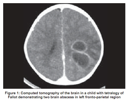
Code Number: ni09064 PMID: 19439863 DOI: 10.4103/0028-3886.51303 Sir, A two-year-old boy, a known case of tetralogy of Fallot (TOF), presented with fever and projectile vomiting of 15 days duration. There was no history of seizures, ear discharge, or altered sensorium. No history of contact with tuberculosis. On examination, his vital signs were stable. His weight was 10kg and height was 85cm. He was plethoric with central cyanosis and grade II clubbing. He had a BCG scar. Cardiovascular system examination revealed a single second heart sound and ejection systolic murmur in the pulmonary area. Ocular fundi were normal. Neurological examination was otherwise normal, except for neck stiffness and bilateral plantar extensor. Investigations revealed: Hemoglobin 15.8gm/dL, total WBC count 15600 cells cu.mm with differential of neutrophils 68% and lymphocytes 25%. Mantoux test and HIV ELISA were negative. Echocardiogram did not reveal any vegetation. Chest X-ray was within normal limits. CT brain demonstrated two brain abscess measuring 3 x 3.5 cm in the left fronto-parietal region with perilesional edema and mass effect [Figure - 1]. A left parietal burr hole was made and 15 ml of pus was aspirated from a depth of 1.5 cm from the cortical surface. Cefotaxime, metronidazole, and cloxacillin were instituted initially. Gram stain of pus did not reveal any organism and aerobic culture was sterile. Zeil Neilson smear did not show acid fast bacilli. He was treated with parenteral antibiotics for two weeks and discharged on oral cloxacillin after he improved symptomatically. Surprisingly, after six weeks, his pus grew mycobacterium tuberculosis sensitive to isoniazid, rifampicin, pyrazinamide, and ethambutol. He was started on isoniazid, rifampicin, pyrazinamide, and ethambutol. Unfortunately, he was lost to follow up after this and the outcome of the illness is not known. Brain abscess in patients with cyanotic heart disease constitutes 11% of all brain abscesses and the incidence is in decline in developed countries due to early surgical interventions in patients with congenital heart diseases.[1] Tuberculoma of brain is an extremely uncommon lesion in patients with cyanotic heart diease. Moorthy et al ., [2] reported brainstem tuberculoma in an adult patient with cyanotic heart disease Ray et al ., [3] reported tuberculoma in a child with congenital heart diseases. Probably this is the first report of tuberculous brain abscess in a patient with cyanotic heart disease. Antituberculous therapy and surgical intervention are the mainstays of management. We report this case since physicians treating children with congenital heart lesions should be aware of this rare association and consider when there is inadequate response to initial broad-spectrum antibiotics. References
Copyright 2009 - Neurology India The following images related to this document are available:Photo images[ni09064f1.jpg] |
| |||||||||

