 |
| About Bioline | All Journals | Testimonials | Membership | News |
|
||||||
|
||||||
Neurology India, Vol. 57, No. 2, March-April, 2009, pp. 225-226 Letter To Editor Parenchymal brain cysts in Schimmelpenning-Feuerstein-Mims syndrome Mahesh Kamate, Arun Dumale, Virupaxi Hattiholi 1 Departments of Pediatrics and 1 Radiology, KLE University’s J N Medical College, Belgaum, Karnataka State, India. Correspondence Address: Department of Pediatrics, KLE University's J N Medical College, Belgaum, Karnataka State, India drmaheshkamate@gmail.com Date of Acceptance: 25-Jan-2009
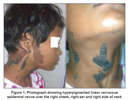
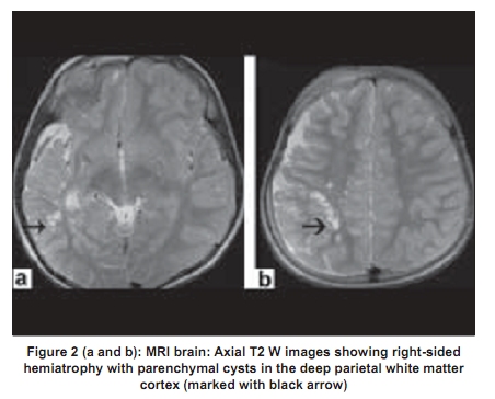
Code Number: ni09071 PMID: 19439869 DOI: 10.4103/0028-3886.51310 Sir, A 10-year-old girl presented with verrucous epidermal nevus over right half of the face and neck [Figure - 1], recurrent complex-partial seizures from six years of age, and poor scholastic performance. The skin lesions evolved with age from pink to yellowish-brown color. She also had limbal dermoid and the rest of the examination including skeletal system was normal. Magnetic resonance imaging (MRI) of the brain revealed right-sided hemiatrophy with parenchymal cysts in the deep parietal white matter [Figure - 2]. The combination of characteristic verrucous skin lesions, ocular findings and the hemiatrophy on MRI are diagnostic of Schimmelpenning-Feuerstein-Mims syndrome (SFM) syndrome. SFM syndrome is a rare neurocutaneous disorder with clinical heterogeneity. The disorder consists of a triad of linear sebaceous nevus, seizures and mental retardation. Central nervous system (CNS) involvement occurs in the form of hemimegalencephaly with ipsilateral gyral malformations, seizures, hemiparesis, mental retardation, cranial nerve palsies and hydrocephalus. [1] Neuroimaging findings in SFM syndrome include the primary lesions: Malformations of cortical development like hemimegalencephaly, polymicrogyria; gliomatosis, hemiatrophy with or without parenchymal cysts, vascular malformations and intracranial/intraspinal lipomas and secondary features: Porencephaly, infarcts and atrophy. [2] Other rare findings include enlarged ventricles, cortical hypoplasia, intracerebral calcification, arachnoid cysts, intracranial tumors and skull asymmetry. [3] CNS complications are more likely to be associated with epidermal nevi on the head and the CNS abnormalities are most often ipsilateral to the skin lesions. [4] Though hemiatrophy has been reported before in SFM, parenchymal cysts in brain have rarely been reported in SFM syndrome. There is mention of porencephalic cysts in the literature [4] but no report of parenchymal cysts like the ones seen in our case. References
Copyright 2009 - Neurology India The following images related to this document are available:Photo images[ni09071f1.jpg] [ni09071f2.jpg] |
| |||||||||
