 |
| About Bioline | All Journals | Testimonials | Membership | News |
|
||||||
|
||||||
Neurology India, Vol. 57, No. 3, May-June, 2009, pp. 350-351 Letter To Editor Multiple giant cavernous angiomas of the brain L. N. Tripathy, S. N. Singh Apollo Gleneagles Hospitals, Kolkata, India. 58, Canal Circular Road, Kolkata - 700 054
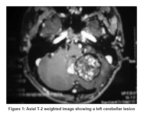
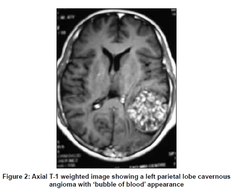
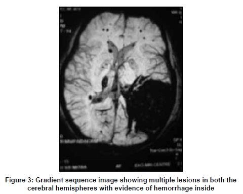
Correspondence Address: Apollo Gleneagles Hospitals, Kolkata, India. 58, Canal Circular Road, Kolkata - 700 054 Date of Acceptance: 07-Mar-2009 Code Number: ni09101 PMID: 19587484 DOI: 10.4103/0028-3886.53267 Sir, Cavernous angioma (CA) of central nervous system (CNS) is an uncommon disease and presents with seizures, hemorrhagic episodes, and rarely with focal deficits. [1] Multiple CAs are often familial. Giant CAs (GCAs) are extremely rare and multiple GCAs are sill rarer. Sometimes, these lesions can be fatal from massive hemorrhage. Most GCAs present as multicystic lesions with hemosiderin ring around on MRI giving a 'bubbles of blood' appearance. [2] Radiofrequency thermocoagulation-assisted surgery [3] and neuronavigation [4] have been advocated by some to treat such lesions with minimal blood loss and precision, respectively. We report a case with multiple CAs with two of the lesions assuming a giant size (more than 4 cm in diameter). A 46-year-old male, was operated for a left cerebellar CA in 1984, a ventriculoperitoneal shunt followed by total excision of the lesion was done. Postoperatively he had improved completely. Follow-up brain computerized tomography (CT) in 1991 showed a left parietal CA. As he was asymptomatic, surgery was not advised. Subsequently, the patient was lost to follow up. In the present admission he was admitted for severe headache, vomiting and ataxia. Magenetic resonance imaging (MRI) of brain revealed a giant cavernoma measuring 5.3 cm in diameter in the left parietal lobe [Figure - 1] and another slightly smaller recurrent lesion in the left cerebellar hemisphere measuring 4.2 cm in diameter [Figure - 2]. There were in addition multiple small lesions in both the cerebral hemispheres with evidence of minor hemorrhages [Figure - 3]. There was no history of similar illness in the family. Through a combined approach (posterior fossa and supratentorial parieto-occipital craniotomy), both these lesions were excised completely by standard microsurgical techniques. The patient had an uneventful postoperative recovery. A follow-up CT scan revealed complete excision of the two giant CAs. The diagnosis of CA was confirmed by histopathological examination. At the time of discharge, he had minimal ataxia. References
Copyright 2009 - Neurology India The following images related to this document are available:Photo images[ni09101f2.jpg] [ni09101f3.jpg] [ni09101f1.jpg] |
| |||||||||
