 |
| About Bioline | All Journals | Testimonials | Membership | News |
|
||||||
|
||||||
Neurology India, Vol. 57, No. 3, May-June, 2009, pp. 351-352 Letter To Editor Post-traumatic isolated superior rectus hematoma Prasad Krishnan, K. Sridhar, Mukul Mondal Department of Neurosurgery, National Neurosciences Centre, Calcutta, India.
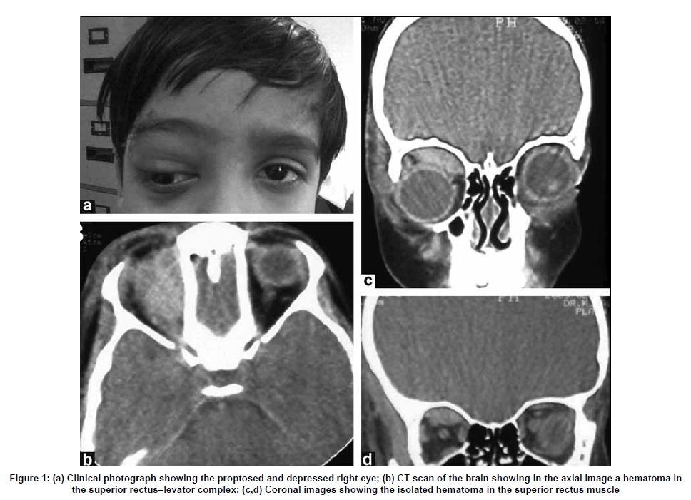
Correspondence Address: Department of Neurosurgery, National Neurosciences Centre, Calcutta, India. Date of Acceptance: 12-Mar-2009 Code Number: ni09102 PMID: 19587485 DOI: 10.4103/0028-3886.53268 Sir, An eight-year-old boy presented with post-traumatic right-sided periorbital ecchymosis and swelling of the eyelid with palpebral fissure closure. A few days later he was unable to fully open the eye and was complained of diplopia. On examination, he had normal visual acuity and fields. The right eye was mildly proptosed and depressed, with periorbital ecchymosis and swelling [Figure - 1]a. There was no subconjunctival or scleral hemorrhage. Fundoscopic examination was normal. Extraocular movements were normal in the left eye. He was able to move the right eye in all directions except superiorly. Forced duction test of the right eye was negative. A CT scan of the brain and orbits showed a hematoma in the superior rectus-levator complex [Figure - 1]b-d. No fracture was seen. The patient was treated conservatively and eye movements gradually improved. Isolated hemorrhage into an ocular muscle following trauma is very uncommon. Only one case of isolated post-traumatic superior rectus hematoma has been reported in the English literature to date. [1] More frequent are reports of inferior rectus hematoma. The commonest cause of a post-traumatic diplopia is mechanical entrapment of soft tissues due to a blow out of the orbital floor. Hemorrhage and edema in the orbital fat causing septae to become taut, can also restrict ocular movement. In both of these conditions, forced duction test is positive. However, in injury to the ocular muscles, the forced duction test is negative. [2] Isolated hemorrhage into ocular muscles does not have any other characteristic clinical feature to distinguish them from other causes of post-traumatic diplopia. Evidence of a bleed may be seen, if the hematoma extends along the muscle sheath to its insertion on the globe. [3] The paucity of clinical findings to distinguish this rare phenomenon from the more common causes of post-traumatic diplopia and proptosis, may result in a wrong or a missed diagnosis. CT scan of the orbit, especially the ++ coronal images, is diagnostic, as it clearly displays the hematoma in the muscle plane. References
Copyright 2009 - Neurology India The following images related to this document are available:Photo images[ni09102f1.jpg] |
| |||||||||
